An algorithmic approach towards the orthoplastic management of osseous and soft tissue defects in post-traumatic distal tibial fractures
2 Orthopaedic Specialists of Northwest Indiana. Volunteer Assistant Clinical Professor, Department of, Indiana University, School of Medicine/Northwest-Gary, Indiana, USA
3 Medical Student, Avalon University School of Medicine, Chief Clinical Rotating Student at Adventist Hospital, Hinsdale IL. Research Associate, Chicago Foot and Ankle Deformity Correction Center, Chicago, USA, Email: HamidKhan2@gmail.com
Received: 01-Jun-2017 Accepted Date: Aug 04, 2017 ; Published: 07-Aug-2017
This open-access article is distributed under the terms of the Creative Commons Attribution Non-Commercial License (CC BY-NC) (http://creativecommons.org/licenses/by-nc/4.0/), which permits reuse, distribution and reproduction of the article, provided that the original work is properly cited and the reuse is restricted to noncommercial purposes. For commercial reuse, contact reprints@pulsus.com
Abstract
Post traumatic fractures involving the lower extremity are often difficult to manage because of the limited options for soft tissue coverage and the increased susceptibility for infection to ensue. One particular anatomic landmark that poses such difficulties is the distal tibia. Currently, there is very limited literature regarding a universal treatment protocol and outcomes related to such defects. This review focuses on some of the promising methods used to describe and define a systematic orthoplastic approach towards diagnosing, managing and treating both osseous and soft tissue defects involved with post traumatic distal tibial fractures.
Keywords
Tibial fractures, Defects, Plastics, Surgical flaps, Algorithms
Introduction
As the field of orthoplastic-microsurgery continues to evolve so do the methods of management and treatment options for patients suffering from distal extremity injuries. In the past, many fractures especially open fractures were considered life threatening because of the numerous complications that accompanied the injury, but thanks to the advances made in orthoplastic microsurgery, those risks have declined dramatically [1]. The protocol aimed at assessing the overall extent of injury and management of a posttraumatic tibial fracture is vital for avoiding amputation as well as evading further complications caused by the fracture. These complications often include additional trauma, acute or chronic infection and as previously stated, the need for limb amputation [2]. Today, Limb salvation in cases of post-traumatic injuries to bone continues to be a challenge for many surgeons. The decision for salvage versus primary amputation consistently presents a strain in determining the best outcome for patients as there are currently no definitive established guidelines to inform treatment decisions, and it is not clear who benefits the most from an early amputation versus attempted limb salvage [3].
Although there is a very limited amount of new data regarding the epidemiology of post-traumatic distal tibial fractures, numerous studies have found that among all the long bones in the body, the tibia is the most commonly fractured [4]. The National Center for Health Statistics cites that there is an average of 492,000 tibial fractures reported every year in the United States [5]. It was also found that nearly 500,000 people undergo treatment for bone defects with a cost of $2.5 billion annually, these numbers are expected to double by 2020 [6]. With figures like this, it is no wonder why osseous and soft tissue defects in post-traumatic distal tibial fractures seem to be occurring more frequently and the need for an ideal orthoplastic protocol to follow is compulsory. Apart from the figures, there are also many doubts related to current surgical recommendations and practices. For instance, many surgeons previously relied on the so called ‘reconstruction ladder’ [7] when it came to making decisions related to an orthoplastic approach towards reconstructive soft tissue options, however, a recent review written by the well-known Fu-Chan Wei questions whether its practice still holds value [8]. These debates and changes in protocol assessment are further proof for the importance of our review and its impact on orthoplastic surgical measures.
It is also important to note that this review focuses mainly on those high energy lower extremity injuries that are commonly categorized as being type IIIB (exposed bone with periosteal stripping and or extensive contamination) and type IIIC (IIIB with additional vascular injury) in association with the Gustilo-Anderson fracture classification [9]. With that said, we feel as though an algorithmic osteoplastic approach towards solving defects of osseous and soft tissue structures in post-traumatic distal tibial fractures becomes monumental for surgeons. The hope of this approach is to eliminate the confusion that is regularly associated with the plan and care of such conditions by enforcing the establishment of an early plan as well as decreasing any likelihood of further complications to transpire.
Applying the Algorithm
For the prepared algorithms to be successful, a systematic approach must be practiced in a step by step fashion. The following discussion will be presented in two separate conditions related to post-traumatic distal tibial fractures. The first being osseous defects and the second being soft tissue defects. The importance of following the algorithm in the proposed structured manner is essential in discovering the greatest advantageous plan of attack towards the management and treatment of osseous and soft tissue defects in post-traumatic distal tibial fractures.
Osseous defects in post-traumatic distal tibia fractures
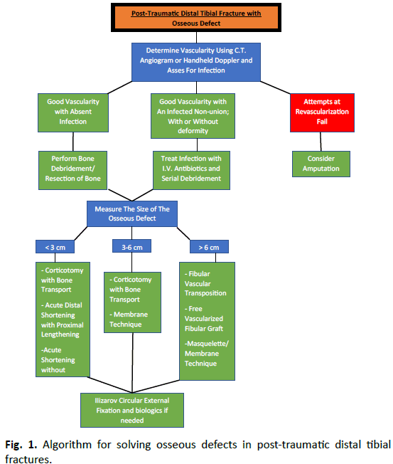
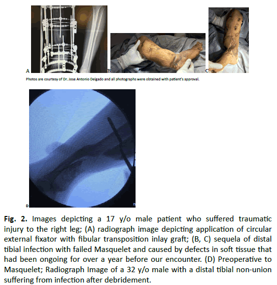
Osseous defects in post-traumatic distal tibia fractures have been associated with numerous deficits and complications, most of which often require the practice of surgical techniques and methods for further management or treatment and the answer to the best means of treatment has yet to be determined [10]. The following algorithm (Fig. 1) depicts the process implemented when evaluating any patient suffering from osseous defects in post-traumatic distal tibial fracture. For this review, distal tibial osseous deficits (Fig. 2) are categorized as being traumatic infected or nonunion defects, with or without deformities. The variant forms of distal tibial osseous deficits involved determine which technique and treatment options are optimal for that circumstance.
Fig. 2. Images depicting a 17 y/o male patient who suffered traumatic injury to the right leg; (A) radiograph image depicting application of circular external fixator with fibular transposition inlay graft; (B, C) sequela of distal tibial infection with failed Masquelet and caused by defects in soft tissue that had been ongoing for over a year before our encounter. (D) Preoperative to Masquelet; Radiograph Image of a 32 y/o male with a distal tibial non-union suffering from infection after debridement.
Examination of vascularity and extent of injury
As with most post traumatic injuries, vascularity and the extent of osseous defect present are key factors in determining what methods of management and treatment options are best for the different defects present in post-traumatic tibial fractures [11]. By using a handheld doppler ultrasound (HDU) and C.T. angiogram we can successfully identify perforators within the lower extremity to help assess vascularity. The use of HDU has been widely used for the preparation and plan of various fascial cutaneous flaps and to distinguish the viability of specific incisions [12]. Once vascularity has been determined, the next step in management is evaluating for infection. In cases where infection does exist, serial debridement and the administration of I.V. antibiotics are necessary until the infected site is eradicated and removal of all nonviable bone or tissue should be performed [13].
When trying to determine vascular regulation, the importance of staging plays an integral part in planning the most appropriate course of action to take. After investigating for signs of venous congestion and an evaluation of choke vessels is completed, a better understanding of the patient’s vascular status can be made [14]. The status of active choke vessels is imperative when deciding if a skin flap will be able to maintain its viability, this topic is discussed in further when we define ‘delay phenomenon’. But to create a quick and concise understanding of the algorithm and for the simplistic purpose of this text, vascular staging should be detected by the presence or absence of any form of a compromised vascular status. In those instances where vascularity has been compromised and C.T. angiogram or HDU findings are not promising, possibilities for revascularization must be evaluated. Poor vascularity can be detrimental to the healing process and all affected patients should be assessed by either an interventional cardiologist, vascular surgeon, or both. If the consensus reached is that revascularization is not possible and that the osseous and or soft tissue defects in the post-traumatic tibial fracture will likely involve a cascade of infection then amputation must be at the top of the list for surgical options [15]. When dealing with cases where infection is controlled, eradicated or absent, debridement of nonviable bone must also be performed. Current recommendation involves low pressure lavage with large volumes of warm saline to complete the debridement of the bone [16].
After non-viable bone debridement has been completed, a measurement regarding the size of the osseous defect must be documented. Based on our professional opinions and personal experience, it is really the diameter of the osseous defect present within the post-traumatic tibial fracture that should guide the surgeon towards determining what the next best step in management should be, as represented in the accompanying algorithm.
Procedures involving the management and treatment of osseous defects in post-traumatic distal tibial fractures
Fixation techniques for distal tibial fractures are a frequent practice in the setting of post-traumatic long bone injury and can be performed in one of two manners; either by internal fixation via means of plates and screws or by external fixation (with or without minimal internal fixation) [17]. Currently, the most popular form of surgical treatment related to internal fixation for tibial fractures is intramedullary nailing; a method using an intermedullary nail to act as a stable rod dedicated to ensuring that the effected bone stays in proper position during the healing process [18]. Results of a recent retrospective study performed to determine the outcomes of intermedullary nailing for distal tibial fractures found that this technique allowed for acceptable range of motion and alignment to be achieved, making it an appropriate and widely practiced option [19].
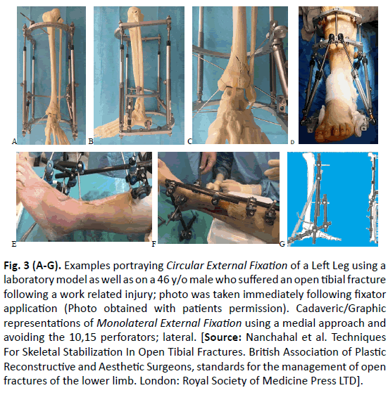
In cases where intermedullary nailing is not considered to be optimal, plates and screws should be implemented to align fragmented bone back to its normal anatomical position or the option for external fixation should be examined [20]. External fixation (Fig. 3) involves the use of pins or screws which are selectively positioned into the bone both above and below the site of the fracture and is performed using one of two methods. These methods comprise of either the use of a circular external technique or a mono-lateral external fixation technique [21]. When implementing external fixation techniques for stabilization in association to local infection, it is important that the technique is positioned away from the infected site to ensure proper healing and to avoid further complications. Studies have shown that based on the severity of the case and the size of defect present, there are various treatment options intended for osseous defects present in post-traumatic tibial fractures [22]. Our algorithm focuses on the use of one or more of the following; Bone grafting, distraction osteogenesis, bone transposition, and the application of the Masquelet technique [23].
Fig. 3 (A-G). Examples portraying Circular External Fixation of a Left Leg using a laboratory model as well as on a 46 y/o male who suffered an open tibial fracture following a work related injury; photo was taken immediately following fixator application (Photo obtained with patients permission). Cadaveric/Graphic representations of Monolateral External Fixation using a medial approach and avoiding the 10,15 perforators; lateral. [Source: Nanchahal et al. Techniques For Skeletal Stabilization In Open Tibial Fractures. British Association of Plastic Reconstructive and Aesthetic Surgeons, standards for the management of open fractures of the lower limb. London: Royal Society of Medicine Press LTD].
Treatment options associated with bone grafting techniques include the use of either an autograft or allograft. Iliac crest tricortical autogenous bone grafts are commonly used and harvested for autologous bone transplantation, this practice continues to remain the gold standard in the treatment of many osseous defects [24]. Bone grafting techniques that involve using allografts should consider using the femoral head. Although there is still a great deal of controversy regarding femoral head bone allografts and its success rate, there have been many cases proving that the method does heal bone and ultimately aids in limb salvation as explained by Santos et al. [25] in a recent study. If dealing with osseous defects greater than 6 cm in diameter, distraction osteogenesis can be an ideal practice for treatment. The method was first described by professor Llizarov during the mid-19th century, for the treatment of bone defects and involves de novo production of bone between divided bone surfaces (corticotomy) undergoing gradual distraction [26]. Distraction osteogenesis either comprises the use of bone transport, where a free fibular transfer from the contralateral leg may be indicated or it can involve the implementation of tibial lengthening techniques. In cases where fibular transposition deems a positive result, the decision for use of a free vascularized fibula versus a distally based vascularized fibula must be calculated in hopes of attaining the best patient outcome.
As far as deciding on what degree of shortening is best for the patient, there remains to be some deal of confusion as there is no
universal set value. Current literature and studies suggest that an approximately 1.5 cm leg discrepancy does not affect walking or functioning, it was found that sufficient bone shortening is better than performing lesser shortening to achieve stable wound care and bone union within the range of 25% to the normal tibia [27]. The management of large gaps present in long bone fractures, particularly those of post-traumatic tibial fractures is often problematic. In these instances, using the innovative technique referred to as ‘Masquelet’s technique’ should be performed. This technique is especially promising for gaps that are greater than 6 cm in size and involves the notion of creating induced membrane formation [28]. The procedure is performed in series of stages, requiring the use of bone cement as a spacer in first stage and autologous cancellous bone graft to fill the gap in second stage [29]. Aside from the previously mentioned treatment options for osseous defects in post-traumatic tibial fractures, adjunct treatment involving the use of othobiologics must also be considered as possibilities. The orthobiologic treatment options include the use of bone marrow aspirate (BMA) [30], platelet rich plasma (PRP) [31], and demineralized bone matrix (DBM) [32].
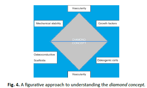
Also, it is important to note that in cases where difficulties to achieve union are expected, application of the ‘diamond concept’ (Fig. 4) may be helpful in providing optimum mechano-biological conditions for bone repair and should be considered [33]. The diamond concept designates five aspects, on which bone healing is based and considered as important pillars for treatment, these include: osteogenic cells (mesenchymal stem cells), osteoinduction (growth factors), osteoconduction (scaffolds), mechanical stability, and vascularization [34]. By assessing each of these concepts in detail, we are able to reach an effective hypothesis as to why the difficulty of achieving union may be occurring. In summary, having a fundamental knowledge of the treatment options for osseous defects in post-traumatic tibial fractures as exhibited in the associated algorithm plays a dynamic part in reaching the best patient outcome and must be addressed prior to taking further surgical action.
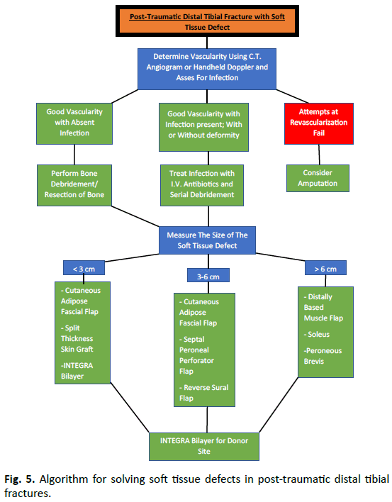
The algorithmic approach for handling soft tissue defects (Fig. 5) in post-traumatic distal tibial fractures follows a similar method to that seen with osseous defects, however, some of management and treatment options create distinguishing factors that allow for a separate discussion and strategy of each scenario to be made. As discussed previously, vascularity and the extent of injury, particularly soft tissue injury for this portion of the review, are major contributors in determining the best methods of management and treatment options for defects related to posttraumatic tibial fractures. HDU and C.T. Angiogram are once again used to determine vascular status and the same steps associated to vascularity and practiced in the osseous defects involved with post-traumatic tibial fracture algorithm are followed. As soon as vascularity has been determined or re-established, the next step in the algorithm is deciding on which orthoplastic soft tissue technique to use. In those instances where vascularity fails to be reestablished, an assessment regarding amputation must be made.
Procedures involving the management and treatment of soft tissue defects in post-traumatic distal tibial fractures
For the purpose of this particular review, some of the soft tissue coverage options for post-traumatic distal tibial fractures include local rotation cutaneous technique of the lower leg, cutaneous adipose fascial flaps, muscle flaps and the use of skin grafts as options intended for management [35]. Based on the reviewed literature, past experience, and personal opinions, it is suggested that the decision for which options are best is once again established via defect size.
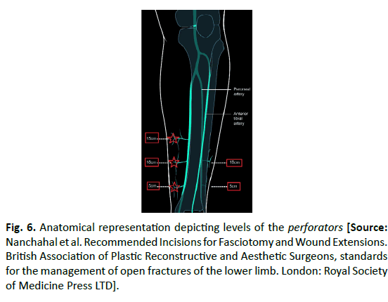
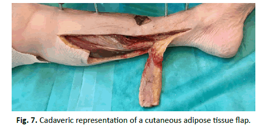
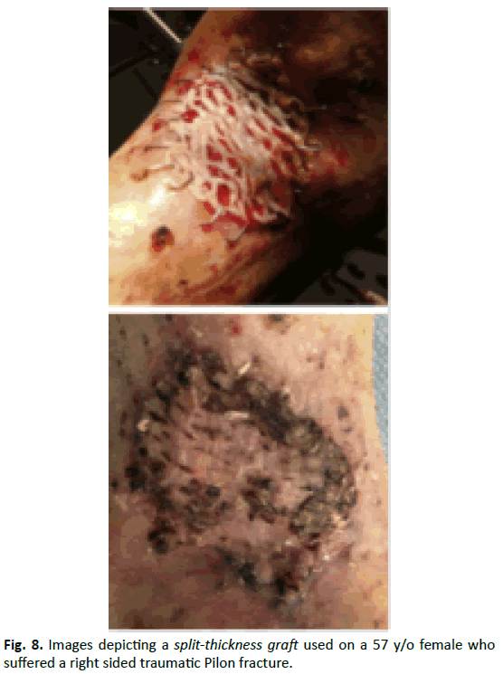
The practice of conventional flaps in relation to extreme injury is rapidly being replaced by perforator flaps and the outcomes related to the use of these flaps as well as the associated benefits continues to develop [36]. One of the many problematic issues that arises while considering the practice of flaps as a means of management is deciding whether a proximal or distally based fascial flap should be used; perforators, choke vessels, angiosomes, and the ‘5,10,15’ concept (Fig. 6) are all principal factors to consider when discussing the use of flaps [37]. The decision for using a cutaneous adipose flap (Fig. 7) has proven to be successful for graft vascularization and integration, two process which are critical in maintaining flap survival [38]. By having a solid understanding of the patient’s circulation and knowing the blood supply of the skin, surgeons lessen the risks associated with wound complications [39]. For defects measuring less than 3 cm in diameter, the use of a split thickness graft (Fig. 8) or Integra bilayer should be considered.
Fig. 6. Anatomical representation depicting levels of the perforators [Source: Nanchahal et al. Recommended Incisions for Fasciotomy and Wound Extensions. British Association of Plastic Reconstructive and Aesthetic Surgeons, standards for the management of open fractures of the lower limb. London: Royal Society of Medicine Press LTD].
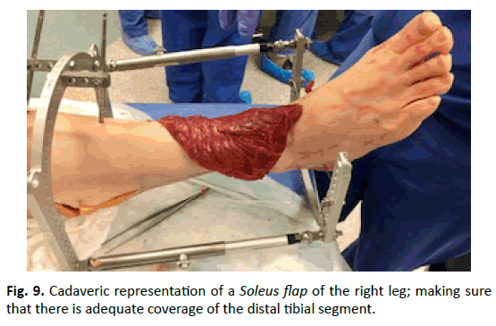
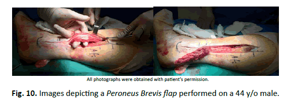
As the size of the soft tissue defect increases, so do the options for flap considerations. Septal peroneal perforator flaps, reverse sural flaps, soleus flaps (Fig. 9) and peroneus brevis flaps (Fig. 10) have all been used as acceptable methods of management for soft tissue defects [40]. After the proper method of management indicated for soft tissue defect has been established, the next course of action is planning treatment. Treatment options for soft tissue defects in post-traumatic tibial fractures are similar to those administered in osseous defects of post-traumatic tibial fractures and orthobiologics should once again be considered in order to reach a thorough assessment of the most superlative treatment route. The orthobiologic options in these instances involve either the administration of an Integra bilayer graft for the donor site or the use of platelets.
Conclusion
There are countless challenges involved with treating defects in post-traumatic distal tibial fractures. As stated previously, the field of orthoplastics and microsurgery is progressing, its practice is essential for many of the advanced cases involving severe post-traumatic injury. Some of the treatment options available include fixation techniques, bone transport, the use of flaps, and orthobiologics. There are also cases where options are very limited due to poor vascularity and amputation must be performed. Regardless of the scenario, the goal of this review was to briefly explain what surgical methods and literature are available in regard to osseous and soft tissue defects in post-traumatic fractures of the distal tibia and supplement that information into an easy Step-bystep algorithm so that a quick yet effective method of management and treatment become applicable. Overtime, as new research develops we are sure that these algorithms may need modification based on the everchanging protocols seen in surgery. Our hope is that this information not only gives insight to the evolution of orthoplastic surgery but that it sets a foundation for guiding surgeons in making the most appropriate plans for care and allows them to feel confident in their decisions.
REFERENCES
- Spyropoulou A., Jeng SF.: Microsurgical coverage reconstruction in upper and lower extremities. Seminars in Plastic Surgery. 2010;24:34-42.
- Nanchahal J., Nayagam S., Khan U., et al.: 15/Open fractures of the foot and ankle. in: standards for the management of open fractures of the lower extremity. London: Royal Society of Medicine Press Ltd; 2009:57-62.
- Busse J.W., Jacobs C.L., Swiontkowski M.F., et al.: Complex limb salvage or early amputation for severe lower-limb injury: A meta-analysis of observational studies. J Orthop Trauma. 2007;21:70-76.
- Court-Brown CM, Rimmer S, Prakash U., et al.: The epidemiology of open long bone fractures Court-Brown. Injury. 1998;29: 529-534.
- Antonova E., Le T.K., Burge R., et al.: Tibia shaft fractures: costly burden of nonunions. BMC Musculoskeletal Disorders. 2013;14:42.
- Pearson J.J., Ong. J., Guda, T.: Translatng biomaterials for bone graft. CRC Press. 2016;127:146.
- Levin L.S.: The reconstructive ladder: An orthoplastic approach. Orthop Clin North Am. 1993;24:393-409.
- Deek N.F., Wei F. It is the time to say good bye to the reconstructive ladder/lift and its variants. Journal of Plastic, Reconstructive & Aesthetic Surgery. 70:539-540.
- Gustilo R.B., Mendoza R.M., Williams D.N.: Problems in the management of type III (severe) open fractures: A new classification of type III open fractures. J Trauma. 1984;24:742-745.
- Giannoudis P.V., Calori G.M., Begue T., et al.: Bone regeneration strategies: Current trends but what the future holds? Injury. 2013;44: S1-S2.
- Alt V., Borgman B., Eicher A., et al.: Effects of recombinant human Bone Morphogenetic Protein-2 (rhBMP-2) in grade III open tibia fractures treated with unreamed nails-A clinical and health-economic analysis. Injury. 2015;46:2267-2272.
- Tashiro K., Harima M., Kato M.: Preoperative color doppler ultrasound assessment in planning of SCIP flaps. J Plast Reconstr Aesthet Surg. 2015;68:979-983.
- Lingaraj R., Santoshi J.A., Devi S., et al.: Predebridement wound culture in open fractures does not predict postoperative wound infection: A pilot study. J Nat Sci Biol Med. 2015;6:S63-S68.
- Mishra S.: A simple method for predicting survival of pedicled skin flaps before completely raising them. Indian Journal of Plastic Surgery. 2011;44:453-457.
- DeCoster T.A., Homedan S.: Amputation osteoplasty. The Iowa Orthopaedic Journal. 2006;26:54-59.
- Nanchahal J., Nayagam S., Khan U., et al.: 16 /Open fractures of the foot and ankle. In: Standards for the management of open fractures of the lower extremity. London: Royal Society of Medicine Press Ltd; 2009:15-9.
- Nanchahal J, Nayagam S, Khan U, et al.: 15/Open fractures of the foot and ankle. In: Standards for the management of open fractures of the lower extremity. London: Royal Society of Medicine Press Ltd; 2009:57-61.
- Talerico M., Ahn J.: Intramedullary nail fixation of distal tibia fractures: Tips and tricks. Journal of Orthopaedic Trauma. 2016;30:7-11.
- Kruppa C., Hoffmann M.F., Sietsema D.L., et al.: Outcomes after intramedullary nailing of distal tibial fractures. Journal of Orthopaedic Trauma. 2015;29:e309-e315.
- Roukis T.S., Kang R.B.: Vascularized pedicled fibula onlay bone graft augmentation for complicated tibiotalocalcaneal arthrodesis with retrograde intramedullary nail fixation: A case series. Journal of Foot and Ankle Surgery. 2016;55:857-867.
- OʼToole R.V., Gary J.L., Reider L., et al.: A prospective randomized trial to assess fixation strategies for severe open tibia fractures: Modern ring external fixators versus internal fixation (FIXIT Study). Journal of orthopaedic trauma. 2017;31:S10-S17.
- Hill C.E.: Does external fixation result in superior ankle function than open reduction internal fixation in the management of adult distal tibial plafond fractures? Foot and Ankle Surgery. 2016;22:146-151.
- Masquelet A.C., Fitoussi F., Begue T., et al.: Reconstruction of the long bones by the induced membrane and spongy autograft. Ann Chir Plast Esthet. 2000;45:346-353.
- Guerado E, Caso E.: Challenges of bone tissue engineering in orthopaedic patients. World Journal of Orthopedics. 2017;8:87-98.
- Godoy-Santos A.L., Amodio D.T., Pires A., et al.: Diabetic limb salvage procedure with bone allograft and free flap transfer: a case report. Diabetic Foot & Ankle. 2017;8:1270076.
- Catagni M.A., Guerreschi F., Lovisetti L.: Distraction osteogenesis for bone repair in the 21st century: lessons learned. Injury. 2011;42:580-586.
- Gulsen M., Ozkan C.: Angular shortening and delayed gradual distraction for the treatment of asymmetrical bone and soft tissue defects of tibia: a case series. J Trauma. 2009;66:E61-E66.
- Masquelet A.C., Begue T. The concept of induced membrane for reconstruction of long bone defects. Orthopedic Clinics of North America. 2010;41:27-37.
- Rigal S., Merloz P., Le Nen D., et al.: Bone transport techniques in posttraumatic bone defects. Orthopaedics & Traumatology: Surgery & Research. 2012;98:103-108.
- Li C., Kilpatrick C.D., Smith S., et al.: Assessment of multipotent mesenchymal stromal cells in bone marrow aspirate from human calcaneus. The Journal of Foot and Ankle Surgery. 2017;56:42-46.
- Angthong C., Khadsongkram A., Angthong W.; Outcomes and quality of life after platelet-rich plasma therapy in patients with recalcitrant hindfoot and ankle diseases: A preliminary report of 12 patients. J Foot Ankle Surg. 2013;52:475-480.
- McAlister J.E., Hyer C.F., Berlet G.C., et al.: Effect of osteogenic progenitor cell concentration on the incidence of foot and ankle fusion. J Foot Ankle Surg. 2015;54:888-891.
- Moghaddam A., Zietzschmann S., Bruckner T., et al.: Treatment of atrophic tibia non-unions according to ‘diamond concept’: Results of one-and two-step treatment. Injury. 2015;46:S39-S50.
- Steinhausen E., Glombitza M., Böhm HJ., et al.: Unfallchirurg. 2013;116:633.
- Kamath J.B., Shetty M.S., Joshua T.V., et al.: Soft tissue coverage in open fractures of tibia. Indian Journal of Orthopaedics. 2012;46:462-469.
- Kim J.T., Kim S.W.: Perforator flap versus conventional flap. Journal of Korean Medical Science. 2015;30:514-522.
- Taylor G.I., Corlett R.J., Dhar S.C., et al.: The anatomical (angiosome) and clinical territories of cutaneous perforating arteries: development of the concept and designing safe flaps. Plastic and reconstructive surgery. 2011;127:1447-1459.
- Freiman A., Shandalov Y., Rosenfeld D., et al.: Engineering vascularized flaps using adipose-derived microvascular EC and MSC. J Tissue Eng Regen Med. 2017.
- Mohd-Yusof N., Ahmad-Alwi A.: Perforators in the leg: why it is important for orthopaedic surgeons to know. Malaysian Orthopaedic Journal. 2017;11:85-87.
- Christopher B.: Reverse Sural Flap with Bifocal Ilizarov technique for tibial osteomyelitis with bone and soft tissue defects. The Journal of Foot and Ankle Surgery. 2016;53:344-349.



 Journal of Orthopaedics Trauma Surgery and Related Research a publication of Polish Society, is a peer-reviewed online journal with quaterly print on demand compilation of issues published.