Comparative study of functional and radiological outcome of augmented external fixator versus volar locking plate in intraarticular fractures of distal end radius
Received: 28-Sep-2022, Manuscript No. jotsrr-22-76179;; Editor assigned: 30-Sep-2022, Pre QC No. jotsrr-22-76179;(PQ); Accepted Date: Oct 23, 2022 ; Reviewed: 14-Oct-2022 QC No. jotsrr-22-76179;(Q); Revised: 17-Oct-2022, Manuscript No. jotsrr-22-76179;(R); Published: 24-Oct-2022, DOI: 10.37532/1897- 2276.2022.17(10).76
This open-access article is distributed under the terms of the Creative Commons Attribution Non-Commercial License (CC BY-NC) (http://creativecommons.org/licenses/by-nc/4.0/), which permits reuse, distribution and reproduction of the article, provided that the original work is properly cited and the reuse is restricted to noncommercial purposes. For commercial reuse, contact reprints@pulsus.com
Abstract
Introduction: Augmented external fixation and Open reduction with volar locking plate are two frequently used modalities in the management of intraarticular fractures of distal end radius. However, there is still controversy regarding the optimal surgical modality. The present study was performed to compare the functional and radiological outcomes ofAugmented External Fixation (AEF) versus Volar Plate Fixation (VPF) in the management of patients with intraarticular fractures of the distal end of the radius.
Materials and Methods: This prospective study was done between December 2019 and December 2021. This study included 40 patients with intraarticular fractures of the distal end of the radius. All patients fulfilling inclusion criteria were randomly allocated into two groups. Group A was treated with an AEF and Group B with VPF. Functional assessment was done by measuring the wrist range of motion, hand grip strength, and Mayo Wrist Score. The radiographic parameters included radial height, radial inclination, and volar tilt. Follow-up was done at 6 weeks, 3 months, and 6 months post-operatively.
Results: In our study at all follow-ups, the VPF group had a significantly better Mayo wrist score and wrist flexion, wrist extension, forearm supination, and pronation compared to the AEF group (p< 0.05). There were no significant differences in terms of hand grip strength and postoperative radiological parameters (p> 0.05).
Conclusion: VPF is a better surgical option as compared to AEF based on our short-term functional outcome in the management of patients with intraarticular fractures of the distal end of radius, on account of better wrist flexion and extension, forearm rotation and Mayo wrist scores, and fewer complication rates.
Keywords
Distal radius fracture, intraarticular, augmented external fixator, volar plate fixation, mayo wrist score
Introduction
Fractures of the distal end of the radius are the most common fractures that present in the department of orthopaedics or emergency [1]. With increasing life expectancy, it is estimated that the incidence of distal end radius fractures will also rise. In elderly individuals, fractures of the distal end of the radius occur most commonly after minor falls (osteoporotic fractures) while in a younger population they are the result of high-energy trauma [2,3]. The distal end of radius fractures can be broadly classified into extra-articular and intra-articular fractures [4]. A vast majority of these fractures are intraarticular and result in disruption of either or both the radiocarpal and radioulnar joints. Anatomical reduction and initiation of early movement remain the main goals of treatment. However, the best method of achieving this remains a debatable topic. Over the last three decades, modalities of treatment have evolved from plaster immobilization to percutaneous wiring and pinning, bridging external fixation to augmented external fixation and in the modern era, internal fixation with various kinds of plating [5-7]. Even with numerous studies being conducted on distal radius fractures, there is only a limited number assessing the comparison of outcomes between modalities of treatment, especially between augmented external fixation and internal fixation with volar plating. Therefore, the present study was conducted to compare closed reduction and augmented external fixation with open reduction and internal fixation using volar locking plates for the treatment of distal radius fractures, in terms of functional and radiological outcomes, and complications.
Materials and Methods
Thisprospective studywas conducted in theDepartment of Orthopaedics of a tertiary care hospital. Institutional ethical committee clearance was obtained before the conduct of the study. Patients presenting with intraarticular fracture of the distal end of the radius were admitted to this hospital. Upon admission, detailed history of the patient was obtained including the mode of the injury. Relevant investigations were done and the patients received primary care.
The inclusion criteria were: the intraarticular distal end of radius fracture, age over 18 years, unilateral distal radius fracture, and no contraindication to anaesthesia. The exclusion criteria were: open fracture, bilateral distal radius fractures, fractures >2 weeks old, any other associated injury/fracture, and pathological fractures.
A total of 40 patients were included in the study. The patients were then randomly divided into two groups of 20 patients each using random numbertables generated online. Group A wastreated with an augmented external fixator (AEF) and Group B with volar plate fixation (VPF). Patients as well asthe next-of-kin were explained about the surgery and written informed consent for the surgery was obtained from all patients.
SURGICAL TECHNIQUE
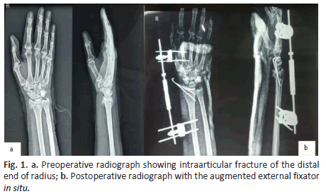
Augmented external fixation: For AEF, two 3.5 mm Schanz pins in the radius proximal to the fracture and two 2.5 mm pins in the second metacarpal were inserted. The Schanz pins were interconnected with connecting rods and universal clamps. After frame application, the reduction was achieved via manual traction (ligamentotaxis) in all cases. Distal fragments were reduced and fixed with the help of two K-wires. Interconnecting rods and clamps were tightened. The final reduction was confirmed under fluoroscopic guidance in anteroposterior and lateral views (Figure 1).
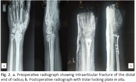
Volar Plate Fixation: Modified Henry’s approach was used to make a longitudinal skin incision. All fracture fragments were reduced and held provisionally with K-wires. Variable angle volar locking plate and screws were placed and position confirmed under fluoroscopic guidance in anteroposterior and lateral views (Figure 2). Postoperatively, a below elbow splint was applied for 4 weeks.
FOLLOW-UP AND POSTOPERATIVE EVALUATION
For both groups, in the early postoperative period, range of motion exercises for shoulder, elbow, and finger joints were started to prevent joint stiffness. In Group A external fixator and K-wires were removed at 6 weeks-8 weeks post-surgery and in Group B below elbow splint was removed at 4 weeks post-surgery and wrist range of motion exercises and grip strength exercises were started. All the patients were followed up at 6 weeks, 3 months, and 6 months postoperatively. Functional assessment was done by measuring the wrist range of motion and hand grip strength. A goniometer was used to measure wrist flexion, extension, supination, and pronation, and hand grip strength was measured using a Jamar dynamometer. All the patients were scored according to the Mayo-Wrist scoring system. Standard anteroposterior and lateral radiographs of the wrist were taken at the final follow-up. Radiological assessment was done by measuring radial height, radial inclination, and volar tilt.
Statistic Analysis
The data were presented in terms of mean ± standard deviation, frequencies, and percentages. To compare categorical variables between the groups, the Chi-square test was used. To compare continuous variables between the groups, the Unpaired t-test was used. To compare the mean change in scores, Paired t-test was used. A p-value of lessthan 0.05 was considered statistically significant.
Results
There were 40 patients in our study, out of which 20 patients underwent AEF and the remaining 20 patients underwent VPF. The demographic attributes of all the patients are present in Table 1.
Table. 1. Patient demographics.
| DEMOGRAPHIC VARIABLES | AEF | VPF |
|---|---|---|
| Age(years) | 51.73 ± 11.09 | 47.70 ± 11.43 |
| Sex(male/female) | Jul-13 | 09-Nov |
| Handedness(dominant/non-dominant) | 12-Aug | 14-Jun |
In our study, most of the patients in both groups were in the age group between 40-60 years. The mean age of patients in the AEF group was 51.73 years, and in the VPF group, it was 47.70 years. In our study, female patients (60%) were more than male patients (40%). The AEF group included 7 males and 13 females and the VPF group included 9 males and 11 females. In our study, the dominant/right wrist (twentysix, 65%) was involved more commonly than the non-dominant/left wrist (fourteen, 35%). In our study, most of the patients had a C2 type fracture (thirteen; 32.5%) followed by C3 (nine; 22.5%), B2 and C1 (six; 15%), and B1 and B3 (three; 7.5%).
The detailed surgery data of the patients are given in Table 2. The mean duration of surgery was 34.33 minutes in AEF group and 75.60 minutes in VPF group. The mean blood loss was 10.20 ml in AEF group and 44.17 ml in VPF group. The differences between the two groups in terms of duration of surgery and blood loss were statistically significant (p<0.05).
Table. 2. Operative record of patients.
| SURGICAL DATA | AEF | VPF | P value |
|---|---|---|---|
| Duration of Surgery (Minutes) | 34.33 ± 4.03 | 75.60 ± 5.24 | <0.0001 |
| Blood Loss (ml) | 10.20 ± 3.03 | 44.17 ± 8.95 | <0.001 |
Functional assessment of all the patients in both groups was done by measuring the wrist range of motion, grip strength, and Mayo wrist scores. The functional outcomes at the final follow-up of 6 months are summarized in Table 3. In our study, wrist range of motion at final follow-up was better in the VPF group when compared to the AEF group and the result was statistically significant. In the VPF group, mean wrist flexion andextension measured 76.33° and 70.13° when compared to 64.22° and 61.37° in the AEF group. Mean supination and pronation in the VPF group measured 75.50° and 74.15° when compared to 64.22° and 61.37° in the AEF group. In our study, grip strength at final follow-up was betterin the VPF group when compared to the AEF group; however, the resultwas not statistically significant. The grip strength was measured 29.77 kg (92% of contralateral) in the VPF group when compared to 26.7 kg (89% of contralateral) in the AEF group. In our study, the Mayo wrist score at the final follow-up was better in the VPF group when compared tothe AEF group and the result was statistically significant. In the VPF group, the mean Mayo wrist score was 84.30 when compared to 77.10 in the AEF group.
Table. 3. Functional outcome at six-month follow-up.
| FUNCTIONAL PARAMETERS | AEF | VPF | P-value |
|---|---|---|---|
| Wrist Flexion(degree) | 64.22±3.71 | 76.33±6.39 | <0.0001 |
| Wrist Extension(degree) | 61.37±2.18 | 70.13±4.33 | <0.0001 |
| Supination(degree) | 69.12±3.46 | 75.50±4.17 | <0.0001 |
| Pronation(degree) | 67.77±3.64 | 74.15±3.73 | <0.0001 |
| Grip Strength(kg) | 26.70±5.3 | 29.77±6.64 | 0.891 |
| Mayo Wrist Score | 77.10±9.7 | 84.30±10.5 | <0.05 |
Radiological assessment was done by measuring radial height, radial inclination, and volar tilt. The radiological parameters at the final follow-up of 6 months are summarized in Table 4. In our study, radiological parameters were restored in both groups. The parameters were better in the VPF group when compared to the AEF group, however, the result was not statistically significant (p>0.05).
Table. 4. Radiological parameters at final follow-up.
| Radiological Parameters | AEF | VPF | P value |
|---|---|---|---|
| Radial height(mm) | 10.5 ± 1.5 | 10.9 ± 1.6 | 0.697 |
| Radial inclination(degree) | 19.7 ± 3.3 | 20.8 ± 4.1 | 0.543 |
| Volar tilt(degree) | 7.5 ± 3.3 | 8.3 ± 3.5 | 0.414 |
In our study, any immediate or late postoperative complications were noted. In the AEF group, 6 cases (30%) of complications occurred. Pin tract infection(3,15%) was the most common complication followed by wrist stiffness (2,10%) and loss of reduction requiring re-adjustment (1,5%). In the VPFgroup, there were 4 cases (20 %) of complications, the most common being scar hypertrophy (2,10%), followed by wrist stiffness (1,5%) and superficial wound infection managed by appropriate antibiotics (1,5%). There was no significant difference in the overall rate of complications between the two groups (p>0.05).
Discussion
Augmented external fixation and Open reduction with volar plating are two frequently used modalities in the management of intraarticular fractures of the distal end of the radius with acceptable functional and radiological results. However, there is still controversy regarding the superior surgical modality. The advantages of augmented external fixation are easy surgical technique, improved reduction by ligamentotaxis, minimal surgical exposure, and less operative time [8]. The advantages of open reduction and volar plate fixation are direct visualization and manipulation of the fracture fragments, stable rigid fixation, and initiation of early wrist motion exercises [9].
In this study, we found that most of the patients in both groups were in the age group of 40 years-60 years. The prevalence in this age group can be attributed to the onset of osteoporosis at this age [10]. There was no significant difference in the gender between the groups showing comparability of the groups in terms of gender, however, a female predominance wasseen in both AEF (65%) and VPF groups(55%). The higher incidence among females could be attributed to post-menopausal osteoporosis. In this study, the dominant side was involved in 26 out of the 40 cases. This could be attributed to a defence mechanism when falling on the dominant side. In this study, we found that the duration of surgery and amount of blood loss was considerably less in the AEF group. These findings were in agreement with observations made by Chaudhary et al. [11].
In this study, we found that wrist range of motion in terms of wrist flexion and extension and forearm supination and pronation were significantly better in the VPF group when compared to the AEF group. This might be attributed to the fact that volar plate fixation allowed an earlier wrist mobilization. These findings corroborate with the results of multiple studies and reviews on the topic done in the past both in the western and Indian populations. A meta-analysis conducted by Cui Z et al. consisted of 738 patients and compared the outcome of external versus internal fixation in intraarticular fracture of the distal radius. The study concluded that the clinical outcome was better in the internal fixation group at a follow-up of six weeks, with similar results at 3 months and 12 months postoperatively, and supported internal fixation over external fixation [12]. Similar results were seen in a meta-analysis conducted by Fu Q et al. that consisted of 776 patients with distal end radius fractures treated with either external fixation or a volar locking plate and concluded that volar plating gives better clinical outcomes even at 12 months of follow-up and hence supported the use of volar plating for the management of distal radius fractures [13]. In this study, there was no significant difference in terms of grip strength between the two groups. However, at all follow-ups, the grip strength was found to be better in patients who underwent volar plate fixation. This can be attributed to the early initiation of movement in the VPF group, which was delayed in the AEF group due to immobilization of the wrist joint with an external fixator. This was comparable to observations made by Abramo et al. and Rozental et al. [14,15].
The Mayo-Wrist Score is a modification of the Geen and O’Brien score [16]. There is a total of 100 points and include four subdomains with 25 points each. The subdomains are pain, wrist dorsiflexion/palmar flexion arc, grip strength, and functional/work status of patients. In this study, we found that the mayo wrist score was significantly higherin the VPF group as compared to the AEF group at all follow-ups of 6 weeks, 3 months, and 6 months. A randomized control trial conducted by Leung F et al. consisted of 137 patients and compared augmented external fixation with volar plate fixation for intraarticular fractures of distal end radius and concluded that at the time of final follow-up, the functional outcome based on mayo wrist scorers was significantly better for the volar plate fixation group than those for the augmented external fixation group [17]. Dwivedi et al. in a similar study found that at the final follow-up, the mayo wrist score of the volar plating group was 85 compared to 78 for the external fixation group, thus concluding that volar plating is a superior surgical modality in terms of functional outcome [18].
In thisstudy, the radiological assessment was done at the final follow-up by measuring radial height, radial inclination, and volar tilt. Acceptable reduction parameters were obtained and maintained at each follow-up in both groups. The parameters were better in the VPF group when compared to the AEF group, however, the result was not statistically significant. Gereli et al., in their study, concluded that, radiographically, the volar plating was associated with better correction of volar angulation [19]. This may be explained by the fact that the subchondral distal screws of the volar locking plate provide support against palmar angulation losses. Wright et al., concluded in their study that radial shortening was prevented in the volar plating group whereas it occurred more in the external fixator group because of relatively poor correction of the articular surface [20].
Limitation
The main limitations of the study include a small sample size, short period of follow-up of six months, and the use of a physician-based scoring system.
Conclusion
Both surgical techniques have efficacy in the management of intraarticular fractures of the distal end of radius but volar locking plate fixation has more advantages over augmented external fixation asshown by early improvement in wrist range of motion, better grip strength and faster return to activities of daily living. Based on our short-term functional outcome, it can be concluded that volar plate fixation is a better surgical option as compared to augmented external fixation in the management of patients with intraarticular fractures of the distal end of radius, on account of better wrist flexion and extension, forearmrotation and Mayo wrist scores, and fewer complication rates.
Acknowledgement
FUNDING
No funding wasreceived for conducting this study
COMPETINGINTERESTS
The authors have no competing interests to declare that are relevant to the content of this article.
ETHICS APPROVAL
Approval was obtained from the ethics committee of Bhabha Atomic Research Centre and Hospital, Mumbai, India.
CONSENT TO PARTICIPATE
Informed consent was obtained from all individual participants included in the study.
CONSENT FOR PUBLICATION
Patients signed informed consent regarding publishing their data.
References
- Graftiaux, A.G., Kehr, P. Y. Zhang: Clinical epidemiology of orthopaedic trauma. Eur J Orthop Surg Traumatol 27, 869 (2017). [Google Scholar] [CrossRef]
- Haus BM, Jupiter JB. Intra-articular fractures of the distal end of the radius in young adults: reexamined as evidence-based and outcomes medicine. J Bone Joint Surg Am. 2009 Dec;91(12):2984-91 [Google Scholar] [CrossRef]
- Jupiter JB, Ring D, Weitzel PP. Surgical treatment of redisplaced fractures of the distal radius in patients older than 60 years. J Hand Surg Am. 2002 Jul;27(4):714-23. [Google Scholar] [CrossRef]
- Xarchas KC, Verettas DA, Kazakos KJ. Classifying fractures of the distal radius: impossible or unnecessary? Review of the literature and proposal of a grouping system. Med Sci Monit. 2009 Mar;15(3):RA67-74. [Google Scholar] [CrossRef]
- Slutsky DJ. External fixation of distal radius fractures. J Hand Surg Am. 2007 Dec;32(10):1624-37 [Google Scholar] [CrossRef]
- Chung KC, Watt AJ, Kotsis SV, et al. Treatment of unstable distal radial fractures with the volar locking plating system. J Bone Joint Surg Am. 2006 Dec;88(12):2687-94 [Google Scholar] [CrossRef]
- Schnall SB, Kim BJ, Abramo A, et al. Fixation of distal radius fractures using a fragment-specific system. Clin Orthop Relat Res. 2006 Apr;445:51-7. [Google Scholar] [CrossRef]
- Handoll HH, Huntley JS, Madhok R. External fixation versus conservative treatment for distal radial fractures in adults. Cochrane Database Syst Rev. 2007 Jul 18;(3):CD006194.[Google Scholar] [CrossRef]
- Kapoor H, Agarwal A, Dhaon BK. Displaced intra-articular fractures of distal radius: a comparative evaluation of results following closed reduction, external fixation and open reduction with internal fixation. Injury. 2000 Mar;31(2):75-9. [Google Scholar] [CrossRef]
- Marwaha RK, Tandon N, Garg MK, et al. Bone health in healthy Indian population aged 50 years and above. Osteoporos Int. 2011 Nov;22(11):2829-36. Epub 2011 Jan 27. [Google Scholar] [CrossRef]
- Chaudhary P, Shrestha BP, Khanal GK, et al. RCT comparing closed reduction and percutaneous versus open reduction and volar locking plate in treatment of distal radius fractures in adults. Int J Orthop Sci 2017;3(2):678-681 [Google Scholar] [CrossRef]
- Cui, Z., Pan, J., Yu, B. et al. Internal versus external fixation for unstable distal radius fractures: an up-to-date meta-analysis. International Orthopaedics (SICOT) 35, 1333-1341 (2011). [Google Scholar] [CrossRef]
- Fu, Q., Zhu, L., Yang, P. et al. Volar Locking Plate versus External Fixation for Distal Radius Fractures: A Meta-analysis of Randomized Controlled Trials. IJOO 52, 602;610 (2018). [Google Scholar] [CrossRef]
- Abramo A, Kopylov P, Geijer M, et al. Open reduction and internal fixation compared to closed reduction and external fixation in distal radial fractures: a randomized study of 50 patients. Acta Orthop. 2009 Aug;80(4):478-85. [Google Scholar] [CrossRef]
- Rozental TD, Blazar PE, Franko OI, et al. Functional outcomes for unstable distal radial fractures treated with open reduction and internal fixation or closed reduction and percutaneous fixation. A prospective randomized trial. J Bone Joint Surg Am. 2009 Aug;91(8):1837-46. [Google Scholar] [CrossRef]
- Green DP, O'Brien ET. Open reduction of carpal dislocations: indications and operative techniques. J Hand Surg Am. 1978 May;3(3):250-65.[Google Scholar] [CrossRef]
- Leung F, Tu YK, Chew WY, Chow SP. Comparison of external and percutaneous pin fixation with plate fixation for intra-articular distal radial fractures. A randomized study. J Bone Joint Surg Am. 2008 Jan;90(1):16-22. [Google Scholar] [CrossRef]
- Dr. Saurabh Dwivedi, Dr. Chandra Prakash Pal, Dr. Kamal Safdar. Comparison of outcome of fracture distal end radius treated by external fixator versus volar plating. Int J Orthop Sci 2020;6(2):715-718. [GoogleScholar] [CrossRef]
- Gereli A, Nalbantol U, Kocao B, et al. Comparison of palmar locking plate and K-wire augmented external fixation for intra-articular and comminuted distal radius fractures. Acta Orthop Traumatol Turc. 2010;44(3):212-9. [Google Scholar] [CrossRef]
- Wright TW, Horodyski M, Smith DW. Functional outcome of unstable distal radius fractures: ORIF with a volar fixed-angle tine plate versus external fixation. J Hand Surg Am. 2005 Mar;30(2):289-99. [Google Scholar] [CrossRef]


 Journal of Orthopaedics Trauma Surgery and Related Research a publication of Polish Society, is a peer-reviewed online journal with quaterly print on demand compilation of issues published.