Fibrolipomatous hamartoma of the sural nerve: An unusual location
Received: 24-Aug-2017 Accepted Date: Sep 14, 2017 ; Published: 18-Sep-2017
This open-access article is distributed under the terms of the Creative Commons Attribution Non-Commercial License (CC BY-NC) (http://creativecommons.org/licenses/by-nc/4.0/), which permits reuse, distribution and reproduction of the article, provided that the original work is properly cited and the reuse is restricted to noncommercial purposes. For commercial reuse, contact reprints@pulsus.com
Abstract
A case of fibrolipomatous hamartoma in the right foot is described here. This lesion commonly occurs in the hands, wrists and forearms of infants, and less commonly in children and young adults. The median nerve is affected in 80% of cases. Only very rarely is it found in the nerves of the foot. It is believed that the present case study is the tenth reported case of fibrolipomatous hamartoma in the foot, and is the first case reported in Nigeria to the authors’ knowledge. The previous nine cases affected the medial plantar nerve in five and superficial peroneal nerve in four. This is the only reported case in English literature affecting a branch of the sural nerve.
Keywords
Foot, Fibrolipomatous hamartoma, Nerve, Neural fibrolipoma
Introduction
Fibrolipomatous hamartoma are benign lesions that occur as soft tissue swelling in relation to a segment of peripheral nerve. They are fusiform swellings that may or may not move across the peripheral nerve depending on the amount of fibrosis surrounding the swelling [1].
It should be noted that the tumour is an anomalous growth of the fibro-adipose tissue of the peripheral nerve sheath [2].
It is commonly found in infants and less commonly in children and young adult [3]. The median nerve is the most affected nerve of the upper limb, followed by the ulna and radial nerve [1]. The foot is rarely affected with just nine cases reported in English literature [2].
A rare case of fibrolipomatous hamartoma of nerve in the foot is described, with emphasis on the unusual location.
Case Report
A 38-year-old woman was seen at our outpatient clinic with a right mid-foot painless swelling noticed about 20 years ago. The swelling has been growing slowly until 2 months prior to presentation when she noticed a more rapid growth. There was no associated neurological symptoms or gait disturbance. No other swelling in other parts of the body. There was no history of trauma, past illness or familial inheritance.
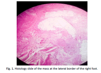
Examination revealed a young woman with a soft, painless 8 × 6 × 4 cm mass at the lateral border of the mid-foot. There was no associated skeletal enlargement or macrodactyly. Radiograph did not reveal any bony lesion. She had excisional biopsy done and a multi-lobulated yellowish mass was found along the distribution of the sural nerve. The tumor was resected with the branch of the nerve. At follow up, wound has healed and no evidence of neurological symptoms. The histopathology slide revealed haphazard arrangement of matured nerve bundles, adipocytes, smooth muscle fibres and fibro-collagenous connective tissues all admixed. Also seen are few vascular channels and areas of hemorrhage as shown in Fig. 1 below.
Discussion
Fibrolipomatous hamartoma has been called by various names, i.e., fibromatosis of nerve, neural fibrolipoma, lipofibroma of nerve, lipomatosis of nerve, and neural lipofibromatosis hamartoma. It has been considered a hamartoma because the fibrous, fatty, and neural components are all essentially mature tissue. Although some consider it to be of congenital origin, the exact etiology remains unclear. It is a fusiform swelling that may or may not move across the peripheral nerve depending on the amount of fibrosis surrounding the swelling. However, it never moves along the nerve. It may be a painful or painless swelling. The histological picture is characteristic; there’s fibrofatty infiltration within the nerve associated with perineural and endoneural fibrosis and thickening of nerve fascicles.
The median nerve is most commonly involved and it is said to account for about 80% of all fibrolipomatous hamartomas, followed by the ulnar, and radial nerve. Lower extremity cases are extremely rare. Fibrolipomatous hamartoma are associated with the overgrowth of bone and macrodactyly in about one-third of cases and are known as macrodystrophia lipomatosa. The differential diagnosis includes ganglion cysts, vascular malformations, traumatic neuroma, and lipomas [1,3-5].
Epidemiologically, fibrolipomatous harmatomas is rare, the first reported case in English literature was in 1953, since then, about 100 cases affecting different parts of the body have been reported, affectation of the foot is very rare [1]. Moreover, indepth knowledge of this tumor was not known in English literature until 1964 and in all, nine histologically confirmed cases of foot fibrolipomatous harmatomas have been reported in the literature. Five of these affected the medial plantar nerve while the remaining four affected the superficial peroneal nerve [2]. The case study here to the best of our knowledge is the tenth case reported in the literature and the only reported case affecting the distribution of the sural nerve. This case study involved a 38 years old woman that has had the lesion for 20 years. That means, it was first noticed when she was 18 years of age and this supported the previous assertion that it affects children [3].
The tumour is a slow growing mass that may have been noticed several years before the patient presents in the hospital. It may be asymptomatic in some patients while in others, it can come with neurological symptoms like pain or paresthesia, physical findings compatible with compression neuropathy or features of carpal tunnel syndrome [3,5].
Magnetic resonance imaging has a characteristic feature that make it the main stay of diagnosis in this lesion. Imaging characteristics are serpiginous low-intensity structures representing thickened nerve fascicles, surrounded by evenly distributed fat, high signal intensity on T1-weighted sequences and low signal intensity on T2- weighted sequences [3,6,7]. In the index case, MRI could not be done.
Microscopically, the lesion is characterized by fibrofatty enlargement of nerve with massive epineural and perineural fibrosis. Mature fat cells have been described within the normal nerve sheath, and it is thought that proliferation of these cells leads to the fatty enlargement of the nerve and its coverings [5].
Conclusion
Excision of the mass can be done although in asymptomatic cases in which diagnosis has been made using MRI, the mass can be left alone [8]. Operative findings are characteristic with the mass looking yellowish and lipomatous in nature and histological diagnosis of the mass should be gotten [9,10]. Index case had excisional biopsy done and histology findings are in keeping with fibrolipomatous hamartoma as shown in Fig. 1.
REFERENCES
- Naik G., Mitra D., Shetty S., et al.: A case report of neural fibrolipoma of foot. J Med Sci Health 2016;2:41-43.
- Ogose A., Hotta T., Higuchi T., et al.: Fibrolipomatous hamartoma in the foot: Magnetic resonance imaging and surgical treatment. JBJS Case Connector. 2002;3:432-436.
- Cavallaro M., Taylor J., Gorman J., et al.: Imaging findings in a patient with fibrolipomatous hamartoma of the median nerve. American Journal of Roentgenology. 1993;161(4):837-838.
- Sureka J., Panwar S., Ahmed M.: Diffuse fibrolipomatous hamartoma of peroneal nerve in the leg, ankle and foot: An uncommon case report. Journal of Musculoskeletal Research. 2011;14:1172001.
- Silverman T., Enzinger F.: Fibrolipomatous hamartoma of nerve: A clinicopathologic analysis of 26 cases. The American journal of surgical pathology. 1985;9(1):7-14.
- Evans H.A., Donnelly L.F., Johnson N.D., et al.: Fibrolipoma of the median nerve: MRI. Clinical Radiology. 1997;52(4):304-307.
- Marom E.M., Helms C.A.: Fibrolipomatous hamartoma: Pathognomonic on MR imaging. Skeletal radiology. 1999;28(5):260-264.
- Maldjian C.: Fibrolipomatous hamartoma of the median nerve on CT. Radiology case reports. 2013;8(4):895.
- Sondergaard G., Mikkelsen S.: Fibrolipomatous hamartoma of the median nerve. The Journal of Hand Surgery: British & European Volume. 1987;12(2):224-225.
- Nodelman L.O., Silverman T.J., Theodoulou M.H.: Neural Fibrolipoma of the Ankle: A case report and review of the literature. The Journal of Foot and Ankle Surgery: Official publication of the American College of Foot and Ankle Surgeons. 2016;55(5):1063-1066.


 Journal of Orthopaedics Trauma Surgery and Related Research a publication of Polish Society, is a peer-reviewed online journal with quaterly print on demand compilation of issues published.