Functional and radiological outcome of unstable distal radius fractures treated with volar plating
2. Department of Orthopaedics, Government District Hospital, Vadakara, Kerala, India
3. Department of Orthopaedics, Medical Trust Hospital, Ernakulam, Kerala, India
4. Department of Orthodontics & Dentofacial orthopedics, Educare Institute of Dental Sciences, Chattiparamba, Malappuram, India
Received: 12-Feb-2022, Manuscript No. jotsrr-22-51926; Editor assigned: 15-Feb-2022, Pre QC No. jotsrr-22-51926(Q); Accepted Date: Feb 27, 2022 ; Reviewed: 18-Feb-2022 QC No. jotsrr-22-51926(QC); Revised: 25-Feb-2022, Manuscript No. jotsrr-22-51926(R); Published: 28-Feb-2022, DOI: 10.37532/1897-2276.2022.17(1).66
This open-access article is distributed under the terms of the Creative Commons Attribution Non-Commercial License (CC BY-NC) (http://creativecommons.org/licenses/by-nc/4.0/), which permits reuse, distribution and reproduction of the article, provided that the original work is properly cited and the reuse is restricted to noncommercial purposes. For commercial reuse, contact reprints@pulsus.com
Abstract
Objective: The purpose of the study was to assess the functional and radiological outcome following surgical management of unstable distal end radius fractures with volar T- plating.
Methods: In this study 25 patients with unstable fracture of the distal end of radius satisfying the inclusion criteria were treated surgically by ORIF with a volar T plate. Patient follow-up was done at 1, 4 and 8 months to assess outcome radiologically and clinically on the basis of range of movements of the wrist, grip strength, and modified Green and O’brien scores. A detailed analysis of complications was also performed.
Results: Results were graded as excellent, good, fair and poor on the basis of modified Green and O’brien score. Out of total 25 patients, 16 patients (64%) achieved an excellent score, 6 patients (24%) had good outcome, 2 patients (8%) had fair score and one (4%) had poor score. All of them attained good bony union. Two of them had screw penetration into the joint and one had infection at surgical site and resulted in fair to poor results in these patients. No wrist arthritis was reported in this study.
Conclusion: Patients with unstable and dorsally displaced fractures of the distal end of radius treated surgically with volar T plating had excellent to good functional results. But these methods can have complications like screw penetration to joint, extensor tendon irritation, infection etc.
Discussion: In present study, TAD was recorded with range of 19 mm to 28 mm with a mean of 22.78 mm. Using Salvati Wilson score at six months based on pain, walking, muscle power and motion and function there were 26% excellent, 52% good, 20% fair and 2% poor cases were identified in our follow up of the fractures treated with sliding hip screw and barrel plate assembly osteosynthesis.
Keywords
Grip strength, T-plate, modified Green and O’brien score, unstable distal radius fractures, volar plating
Introduction
Fractures of the distal end of radius have been discussed in Orthopedic literature in the last 200 years [1]. The fracture patterns were described even before the advent of x-rays. Fractures of distal end of radius are most common fractures of the upper extremity and constitute 75% of all forearm fractures and 17 % of all fractures [2]. Common practice in olden days of treating these fractures has been closed manipulation and reduction followed by immobilization in plaster cast [3,4]. In 1960, Sir John Charnley wrote: ‘It is very fortunate thing that excellent functional results usually follow the common Colle’s fracture, because disappointing results occasionally develop even in the most skillful hands [3]. But in more complex fractures of distal radius patients may not enjoy perfect freedom in all wrist movements and they may not exempt from pain even after many months. Sometimes they end up in malunion of fracture and subluxation /dislocation of distal radioulnar joint resulting in poor functional and cosmetic result [5]. As open reduction and volar plating ensures good results in displaced fractures reduction and its maintenances [6,7]. Hence in this study we evaluate the functional and radiological outcome of surgically managed displaced distal end radius fractures treated with volar T-plate fixation.
Among many classifications systems available for distal end radius fracture; no classification was adequate due to the broad spectrum of injuries and large number of variables to consider. Most of these classification systems are based on number of intra-articular fragments, location of fracture, direction of displacement and involvement of ulna. A good classification should categories the fracture type and the injury severity to guide treatment. AO classification for distal end radius fracture to document any ulnar sided involvement, sub classifies volar distal radius fractures more accurately were used in this study [8].
Materials and Methods
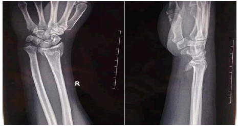
This study was conducted in the Department of Orthopaedics, Government Medical College Trivandrum during the time period between February 2018 and January 2019; which is designed as a prospective case series study of 25 patients with follow up period of 8 months. The study was conducted on patients who were diagnosed as having unstable distal radius fracture and managed surgically by open reduction and internal fixation with Ellis T plate. AO classifications for distal end radius fracture were used; out of 25 patients 3 were C1 fracture, 9 had C2 fracture and 13 patients had C3 facture. The patients coming with this particular fracture was first subjected to closed manipulative reduction and immobilized in long arm slab followed by check x ray. Study inclusion criteria constitutes patients with displaced distal end radius fracture and failed closed manipulation reduction requiring surgical management, which is defined radiologically as >15º of dorsal angulation, >2 mm of articular step-off or gap, or >5 mm of radial shortening, and required surgical fixation and gave consent to participate in the study were included (Figure1). Exclusion criteria constitutes patients with comorbid conditions preventing surgical intervention, patients with immature skeleton, patients with poor skin conditions, patients with more than 3 weeks duration of injury, patients who didn’t give consent to participate in the study.
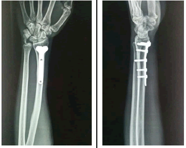
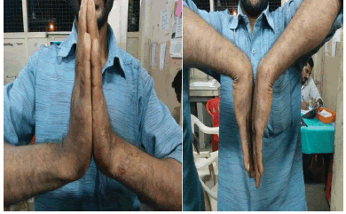
All fractures were approached through Ellis’s approach and fracture was reduced anatomically followed by fixation using volarly placed T-plate [9]. Postoperative x-rays then taken to evaluate the reduction. During follow up visits of 1 month, 4 months and 8 months radiological assessments were done using serial x-rays and evaluated for fracture union, volar angulation (palmar tilt), radial inclination, radio carpal alignment, ulnar variance and articular step and arthritic changes (Figure 2) [11-14]. Patients were evaluated for clinical parameters during each follow-up visits at outpatient clinics regarding pain, residual deformity, palmar flexion, dorsiflexion, supination, pronation and loss of grip strength. (Figure 3). On final follow up at 8 months the patient’s functional status was assessed with mayo wrist score10 which is Modified Clinical Scoring System of Green and O’Brien [11].
DATA ANALYSIS
The data were collected while the patients were admitted in the ward and also at the time of post-operative follow up in OPD at 1st, 4th & 8th postoperative month. The data entered in to Microsoft excel programmer and required analysis was done using descriptive statistical analysis (SPSS version 16). Conclusions were made based on this analysis.
Results
When the final outcome was compared to range of wrist movements it showed that all movements were significantly higher in Mayo excellent group compared to good to poor group. Mean dorsiflexion was 63.8º in excellent result group while 53.3º in poor to good. Mean palmar flexion was 70.6º in excellent compared to 61.1º in the other group. Mean ulnar deviation was 34.1º in excellent while 27.8º in poor to good. Radial deviation was 15º in excellent while 10º in poor to good. Mean pronation in excellent result group was 78.1º while 70º in the other group. Mean supination was 750 in excellent while 67.8º in poor to good. P value <0.05 shows statistically significant results in all wrist movements [Table 1].
Table 1. Comparison between wrist movements and outcome
| Outcome | N | Mean | SD | T | p | |
|---|---|---|---|---|---|---|
| Dorsiflexion | Excellent | 16 | 63.8 | 5 | 4.307 | <0.001 |
| Poor to Good | 9 | 53.3 | 7.1 | |||
| Palmarflexion | Excellent | 16 | 70.6 | 7.3 | 3.058 | 0.006 |
| Poor to Good | 9 | 61.1 | 7.8 | |||
| Ulnar deviation | Excellent | 16 | 34.1 | 3.8 | 3.779 | 0.001 |
| Poor to Good | 9 | 27.8 | 4.4 | |||
| Radial deviation | Excellent | 16 | 15 | 2.6 | 4.699 | <0.001 |
| Poor to Good | 9 | 10 | 2.5 | |||
| Pronation | Excellent | 16 | 78.1 | 4 | 2.652 | 0.014 |
| Poor to Good | 9 | 70 | 11.2 | |||
| Supination | Excellent | 16 | 75 | 5.8 | 2.445 | 0.023 |
| Poor to Good | 9 | 67.8 | 9.1 |
Loss of grip strength was calculated with help of hand-held dynamometer. First the grip strength of both sides was measured and then loss of strength of the affected side was calculated as percentage of loss compared to normal side. It was found that in patients with excellent outcome the mean loss of grip strength was only 0.6% with SD of 2.5, while in poor to good group the mean loss was 16.1% with SD of 10.5, which was statistically significant(p<0.05) [Table 2].
Table 2. Comparison between loss of grip strength and outcome
| Outcome | N | LOSS OF GRIP STRENGTH (% compared to contralateral grip) | T | p | |
|---|---|---|---|---|---|
| Mean | SD | ||||
| Excellent | 16 | 0.6 | 2.5 | 5.686 | <0.001 |
| Poor to Good | 9 | 16.1 | 10.5 | ||
When comparing type of fracture and functional outcome; out of total 12 cases of C1 and C2 fractures 11 patients came out with excellent score while only one patient got a good score. But out of the total 13 C3 fractures, 8 patients got poor to good score and only 5 patients got excellent outcome. Chi-square test analysis showed statistically significant difference in results [Table 3].
Table 3. Comparison between fracture type and outcome
| Fracture classification | Outcome | Total | ||||
|---|---|---|---|---|---|---|
| Excellent | Poor to good | |||||
| N | % | N | % | N | % | |
| C1/C2 | 11 | 68.8 | 1 | 11.1 | 12 | 48.00% |
| C3 | 5 | 31.3 | 8 | 88.9 | 13 | 52.00% |
| Total | 16 | 100 | 9 | 100 | 25 | 100 |
When comparing complications and outcome; it was found that 55% of patients in the poor to good outcome group were associated with some sort of complication while only 6% of patients in the excellent outcome group were associated with complication [Table 4].
Table 4. Comparison between complications and outcome
| Complications | Outcome | Total | ||||
|---|---|---|---|---|---|---|
| Excellent | Poor to good | |||||
| N | % | N | % | N | % | |
| Nil | 15 | 94 | 4 | 45 | 19 | 76 |
| Infection | 0 | 0 | 2 | 22 | 2 | 8 |
| Impingement on tendons | 1 | 6 | 0 | 0 | 1 | 4 |
| Screw penetration to wrist joint | 0 | 0 | 2 | 22 | 2 | 8 |
| Pain over implant | 0 | 0 | 1 | 11 | 1 | 4 |
| Total | 16 | 100 | 9 | 100 | 25 | 100 |
Discussion
The aim of this prospective study was to assess functional and radiological outcome of unstable distal end radius fractures treated surgically with volar T-plating. Those patients satisfying the inclusion criteria were evaluated clinically and radiologically. They were then managed surgically and further evaluated and followed up using particular questionnaire, clinical methods and radiography for a period of up to 8 months. Using modified Green and Obrien’s scoring system functional outcome assessment was done.
In our study 80% cases were below 40 years of age. Unstable type of distal end radius fractures usually results following high velocity trauma such as road traffic accidents and fall from height, the usual victims being young adolescents and 3 times more common in males compared to females. Majority of fractures dealt in the study came under AO type C3 (52%) followed by C2 (32%). C1 type fractures were very few probably due to the fact that most of them had an acceptable position on closed manipulation and would have been managed conservatively. C1 and C2 fractures resulted in a better functional outcome compared to C3 fractures.
At the 8th month follow up most of the patients came up with a pain free wrist movement, others with a mild pain except one who developed moderate pain limiting his activities. The cause of the poor result of this particular case was found to be post-operative infection of surgical site. Achieving normal volar tilt is an important feature in regaining normal grip strength. In this study all patients achieved an acceptable volar tilt except one who had a residual dorsal angulation. Final functional outcome of the study during patient follow- up visits in outpatient clinic were assessed using modified Green & Obrien’s scoring system. Majority of patients (88%) had excellent to good score. Three patients (12%) had a fair to poor score. Similar results were obtained in the study by J. Arora et al. published in 2005 [15]. Final functional status of the patients is encouraging. A good range of pain free wrist movement was achieved in majority of patients. The mean palmar flexion was 670 and dorsiflexion was 600. The mean radial deviation was 130 and ulnar deviation was 320. The mean supination was 720 and pronation was 750. Majority of the patients (64%) regained their normal grip strength. In two patients there was 30 % loss of grip strength which affected their daily activities.
In our study all the patients showed adequate bony union and normal radiocarpal alignment at the final follow-up at 8th month. Volar angulation, ulnar angulation and radioulnar variance at the final follow-up were acceptable in all the patients which did not show any influence in the final functional outcome. There was no evidence of osteoarthritis in any patients at the final follow up, may be because of the short-term nature of the study. Majority of patients (76%) in this study had no complications. Very few patients had complications such as infection and screw penetration into the joint in this present study. These two complications affected the wrist movements adversely and resulted in either fair or poor outcome in this study. Loss of reduction, reflex sympathetic dystrophy, extensor pollicis rupture and deep vein thrombosis are the other complications described with volar buttress plating of distal radius, in literatures of Mehra et al and Jupiter et al [16,17]. In this present study we did not encounter these complications in any of our patients.
Conclusion
In this present study to assess the functional and radiological outcome of the distal end radius fracture treated with open reduction and volar T- plating, majority of fractures dealt with was AO type C2 and C3 fractures of the right hand, mostly in young active males below the age of 40. Most common reasons for these types of fractures where due to motor vehicle accidents, fall from heights and sports injury. Open reduction and internal fixation with Ellis T plate in these patients gave 88% excellent to good outcome with bony union in all patients with acceptable radiocarpal alignment. Most of the patients (84%) returned to their regular activities achieving normal radiological indices of the wrist and grip strength. Majority of patients attained good range of pain free wrist movements by 8th month follow up. Open reduction and internal fixation of the unstable distal end of radius fractures using Ellis T plate proved to be a good option producing good results with very few complications such as infections and screw penetration.
Ethical Statement
Institutional ethical committee clearance was taken to conduct study. Patients were informed that data from the research would be submitted for publication and gave their consent.
REFERENCES
- Bucholz R.W., Heckman J.D.: Rockwood Greens Fractures in Adults. Lippincott Williams & Wilkins. 5th edition. 2001. [CrossRef] [Google Scholar]
- Colles A.: On the fracture of the carpal extremity of the radius. Edinburgh Med Surg.1814;10:182.[CrossRef] [Google Scholar]
- Charnley J.: The closed treatment of common fractures. Churchill Livingstone, Edinburgh. 1950.[CrossRef] [Google Scholar]
- Merchan E.C.R., et al.: Plaster cast versus Clyburn external fixation for fractures of the distal radius in patients under 45 years of age. Orthop Rev. 1992;21:1203-1209.[Crossref][Google Scholar]
- Knirk J.L., Jupiter J.B.J.: Intra-articular fractures of the distal end of the radius in young adults. Bone Joint Surg Am. 1986;68:647-659. [Crossref] [Google Scholar]
- Jansky W., et al.: Klinische und radiologische Ergebnisse von handgelenknahen Speichen- brüchen. Unfallchir. 1994;4: 197-202. [Crossref][Google Scholar]
- Frykman G.K.: Fracture of the distal radius including sequelae shoulder hand finger syndrome. Disturbance in the distal radioulnar joint and impairment of nerve function: A clinical and experimental study. Acta Orthop Scand. 1967;38:1-61.[CrossRef] [Google Scholar]
- Ilyas A.M., Jupiter J.B.: Distal radius fractures: classification of treatment and indications for surgery. Hand Clin. 2010;38:167-173.[CrossRef ] [Google Scholar]
- Ellis J.: Smit’s and Barton’s fractures a method of treatment. J Bone Joint Surg Br. 1965;47:724-727. [CrossRef] [Google Scholar]
- Cooney W.P., et al.: Difficult wrist fractures, perilunate fracture dislocations of the wrist. Clin Orthop Relat Res. 1987;214:136-147. [CrossRef] [Google Scholar]
- Green D.P., O’Brien.: Open reduction of carpal dislocations, indications and operative techniques. J Hand Surg Am. 1978;3:250-265.[CrossRef] [ Google Scholar]
- Schulnd F., et al.: A normal data base of posteroanterior roentgenographic measurements of the wrist. J Bone Joint Surgery. 1992;74:1418-1429. .[CrossRef] [Google Scholar]
- Wood M.B., Berquist T.H.: The hand and wrist. Imaging of Orthopedic Trauma. New York, Raven Press. 1992.[CrossRef] [Google Scholar]
- Kenneth A.E., Kenneth J.K., Joseph D.: Zuckerman Hand book of fractures. 4th edition. 1967. [CrossRef] [Google Scholar]
- Arora J., Malik J.C.: External fixation in comminuted, displaced intra articular fractures of the distal radius: is it sufficient?. Arch orthopedic trauma surg. 2005;125:536-540.[CrossRef] [Google Scholar]
- Mehara A.K., et al.: Classification of volar Barton fractures. Injury. 1993;24:55-59.[CrossRef] [Google Scholar]
- Jupiter J.B., et al.: Operative treatment of volar intra-articular fractures of the distal end of the radius. J Bone Joint Surg Am. 1996;78:1817-1828.[CrossRef] [Google Scholar]


 Journal of Orthopaedics Trauma Surgery and Related Research a publication of Polish Society, is a peer-reviewed online journal with quaterly print on demand compilation of issues published.