Functional outcome in patients with extra-articular distal femur treated with retrograde intramedullary nailing versus minimally invasive plate osteosynthesis- A cross-sectional study
2 Department of Traumatology and Surgery, MGM Medical College, Kamothe, Navi-Mumbai, Maharashtra, India, Email: abc@gmail.com
Department of Orthopaedics, MGM Medical College, Kamothe, Navi-Mumbai, Maharashtra
, India, Email: mohit.issrani12@gmail.comReceived: 26-Dec-2020 Accepted Date: Feb 06, 2021 ; Published: 15-Feb-2021, DOI: 10.37532/1897-2276.2021.16(1).8
This open-access article is distributed under the terms of the Creative Commons Attribution Non-Commercial License (CC BY-NC) (http://creativecommons.org/licenses/by-nc/4.0/), which permits reuse, distribution and reproduction of the article, provided that the original work is properly cited and the reuse is restricted to noncommercial purposes. For commercial reuse, contact reprints@pulsus.com
Abstract
Introduction- Treatment of extra-articular distal femur fractures remains challenging. The present study aimed to compare the functional outcome of these fractures treated by retrograde nailing versus minimally invasive plate osteosynthesis. Method- This randomized trial was conducted between March 2018 and Feb 2020 at a tertiary care trauma center in Navi-Mumbai on 71 consecutive patients with extra-articular distal femur fractures. The inclusion criteria were closed extra-articular distal femur fractures occurring in adults. Floating knee injuries, ipsilateral lower limb fractures, pathological fractures, compound, and fractures with neurovascular injuries were excluded from the study. Thirty-seven patients in group A were treated with intramedullary nailing while Thirty-four patients in group B were treated by minimally invasive plate osteosynthesis. All the patients were followed up at 3,6,12 and 24 months respectively. Results- The mean age of the patients was 58.41 ± 4.21and 56.72 ± 8.13 in groups A and B respectively. The most common mechanism of injury was road traffic accidents with 42 (59.15%) cases. The most common fracture type was 33A2. The mean operative duration was 112.34 ± 13.8 minutes in group A and 98.28 ± 9.76 minutes in group B respectively which was statistically significant (p<0.0044). There were 3 (8.1%) cases in group A and 1 (2.9%) cases in group B who had superficial infection. There were 2 (5.4%) cases in group A and 4 (11.7%) cases in group B who had post-op knee stiffness. As per the Schatzker and Lambert criteria, 58 (81.6%) of the cases had excellent and good results. Conclusion- Although intramedullary nailing is associated with shorter operative time, the final results of both nailing and plate osteosynthesis are similar with no implant superior to another. Both the techniques have similar complication rates
Keywords
extra-articular distal femur fractures, intramedullary nailing, plating
Introduction
Distal femur fractures account for less than 1% of all fractures and 4%- 7% amongst all femoral fractures [1]. Supracondylar fractures are the ones that occur within 9 cm of the distal femur. They usually are a result of high-velocity injuries in young adult men, while they can result due to low-velocity injuries in geriatric age groups [2].
The primary goal of treatment is to maintain the mechanical axis of the lower limb, achieve adequate stability with good functional outcome. A strong fixation method is usually mandatory owing to the presence of a large number of muscle forces around the distal femur.
The optimal treatment method of extra-articular distal femur fractures remains controversial and involves numerous intramedullary nailing and plating types [3-7]. Open reduction is often associated with a high rate of non-union and infection. To decrease this, the concept of biological osteosynthesis and a Minimal invasive approach has been developed. Retrograde intramedullary nailing is a ‘biological’ method, which is preferred by some for its good control of the distal fragment [8,9]. Meanwhile, Locked Plating (LP) remains a popular and effective alternative method to treat these challenging injuries, especially in comminuted fractures. The anatomically pre-contoured locked plate provides added advantage using a limited approach [10,11].
The present study aimed to compare the functional outcome in extraarticular distal femur fractures treated using retrograde intramedullary nailing and minimally invasive plate osteosynthesis.
Materials and Methods
Trial design
This cross-sectional study was conducted at a tertiary high volume trauma care center in Navi-Mumbai, Maharashtra, India between March 2018 and Feb 2020.
Participants and randomization
A total of 102 patients presented with a distal femur fracture of which, 74 patients met the inclusion criteria and were initially enrolled in the study. There was one patient who died because of pre-existing comorbidities, one year after the surgery, while was one patient was lost to follow-up and thus both were excluded from the study.
A total of 71 patients were included in the study at the end. All the patients explained in detail about the procedure and randomization was done using the closed opaque envelope method, which was opened just before the surgery. Based on this, the decision of nailing or plating was taken. The patients were divided into two groups. Thirty-seven patients in group A were treated with intramedullary nailing while the thirty-four patients in group B were treated by minimally invasive plate osteosynthesis. The Institutional Ethical Committee approval was obtained before the commencement of the study.
Inclusion and exclusion criteria
All skeletally matured patients with closed extra-articular distal femur fractures were included in the study. Associated fractures of the ipsilateral lower limb, floating knee injuries, pathological fractures, compound fractures, and fractures with neurovascular injuries were excluded from the study.
Surgical technique
All the procedures were performed under spinal anesthesia on a radiolucent table with a bolster beneath the knee to relax the forces of gastrocnemius muscle. Three doses of second-generation cephalosporin (one pre-operatively and two doses postoperatively at an interval of 12 hours was administered).
Intramedullary nailing
After preparing the distal femur and knee, a 4 cm linear infrapatellar approach was used. The knee joint was exposed after incising the patellar tendon vertically. An entry point with an awl was then made, anterior to the Blumensaat’s line in line with the femoral axis. The entry was then expanded with the starting reamer following which the nail was inserted. The distal and proximal locking was then performed over the zig. The patellar tendon was then repaired and the wound was closed over layers.
Distal femur plating
A lateral approach with approximately 4 cm incision starting above the Gerdy’s tubercle proximally was made. The iliotibial band was incised in line with its fibers and vastus lateralis muscle was elevated and indirect reduction of the fracture was done. The bridge plate was then applied and temporarily held with wires after adequate reduction was achieved and checked with fluoroscopy in both the orthogonal views. Proximal screws were inserted through stab incisions. The wound was closed over layers without a drain.
Post-operative protocol
The active-assisted range of motion exercises was begun immediately as per the pain tolerance. Toe touch weight-bearing was started under supervision from postoperative day 2 with the help of crutches. Full weight-bearing was tolerated as per pain tolerance.
Follow-up and outcome analysis
Regular follow-up for all the patients was done at 3,6,12 and 24 months respectively.
The outcome was measured using the Schatzker and Lambert criteria (Table 1).
Table 1. Schatzker and Lambert criteria
Excellent |
Full extension |
| Flexion <100 degrees | |
| No varus/valgus or rotational deformity | |
| No pain | |
| Perfect Joint congruency | |
| Good | Not more than one of the following |
| Length loss >1.2cm | |
| Varus/valgus deformity <10 degrees | |
| Flexion loss >20 degrees | |
| Minimal pain | |
| Moderate | Any two of the criteria in good category |
| Poor | Any of the following |
| Flexion <90 degrees | |
| Varus/valgus >15 degrees | |
| Joint incongruency | |
| Disabling pain |
Statistical analysis
All the categorial qualitative data variables were calculated using an unpaired t-test. The results were expressed as mean with standard deviation and p<0.05 was considered to be statistically significant. The analysis was done using the Epi-info software (Version 3.4.3) and Microsoft Excel 2013 (Microsoft Office v15.0).
Results
The mean age of the patients was 58.41 ± 4.21 and 56.72 ± 8.13 in groups A and B respectively. The most common mechanism of injury in the present study was road traffic accidents with 42 (59.1%) cases. As per the AO-OTA classification system for distal femur fractures, 33A2 fracture type was most common involving 36 (50.7%) cases followed by 33A3 involving 23 (32.3%) cases while 33A1 was the least common type with 12 (17%) cases. The mean operative duration was 112.34 ± 13.8 mins in group A and 98.28 ± 9.76 mins in group B respectively which was statistically significant (p<0.0044). There were 3 (8.1%) cases in group A and 1 (2.9%) cases in group B who had a superficial infection which was managed with oral antibiotics. There was no case with deep infection in the present study. There was no case with nonunion or delayed union in the present study. No case had any coronal or rotational plane deformity. There were 2 (5.4%) cases in group A and 4 (11.7%) cases in group B who had post-operative knee stiffness at 3 months, which were treated with physiotherapy. However, both of them had a residual extension lag of 5 degree and 10 degree respectively (Table 2). No case had shortening in the present study. The outcome as per the Schatzker and Lambert criteria was as shown in Table 3. There were 17 (62.9%) cases with excellent, 7 (25.9%) cases with good, and 3 (11.2%) cases that had moderate results respectively. There were no cases that had poor results in the present study (Table 3).
Table 2. Demographics and Characteristics
| Characteristic | Group A n=37 (%) | Group B n=34 (%) | Test of significance | p value |
|---|---|---|---|---|
| Age | 58.41 ± 4.21 | 56.72 ± 8.13 | Unpaired t-test | 0.4992 |
| Sex | Chi-square | 0.2887 | ||
| Male | 16 (43.3) | 19 (55.8) | ||
| Female | 21 (56.7) | 15 (44.2) | ||
| Mechanism of injury | Chi-square | 0.5907 | ||
| Road traffic accident | 23 (62.2) | 19 (55.8) | ||
| Fall | 14 (37.8) | 15 (44.2) | ||
| Classification | Chi-square | 0.7011 | ||
| 33A1 | 5 (13.5) | 7 (20.5) | ||
| 33A2 | 19 (51.3) | 17 (50) | ||
| 33A3 | 13 (35.2) | 10 (29.5) | ||
| Operative duration (mins) | 112.34 ± 13.8 | 98.28 ± 8.76 | Unpaired t-test | 0.0044 |
| Intra-operative blood loss | 156.34 ± 42.21 | 153.87 ± 45.61 | Unpaired t-test | 0.885 |
| Complications | Chi-square | 0.197 | ||
| Superficial Infection | 3 (8.1) | 1 (2.9) | ||
| Knee stiffness | 2 (5.4) | 4 (11.7) | ||
| Knee range of movements | 103.45 ± 8.13 | 100.23 ± 13.42 | Unpaired t-test | 0.2213 |
| Radiological union (weeks) | 21.16 ± 5.6 | 22.32 ± 6.3 | Unpaired t-test | 0.617 |
Table 3. Final outcome
| Category | Group A n=37 (%) | Group B n=34 (%) |
|---|---|---|
| Excellent | 18 (48.6) | 16 (47) |
| Good | 11 (29.8) | 13 (38.3) |
| Moderate | 6 (16.2) | 4 (11.7) |
| Poor | 2 (5.4) | 1 (3) |
Discussion
Extra-articular distal femur fractures can be treated by conservative as well as operative management. Undisplaced or minimally fractures can be very well managed conservatively. Nonetheless, these treatment methods of prolonged immobilization are often associated with joint stiffness, quadriceps wasting, secondary displacement.
Surgical modalities for managing these injuries can vary from external fixator [12,13] plating [14,15] or intramedullary nailing [16-18]. The external fixators are better suited for undisplaced and compound fractures where the risk of operative intervention is increased.
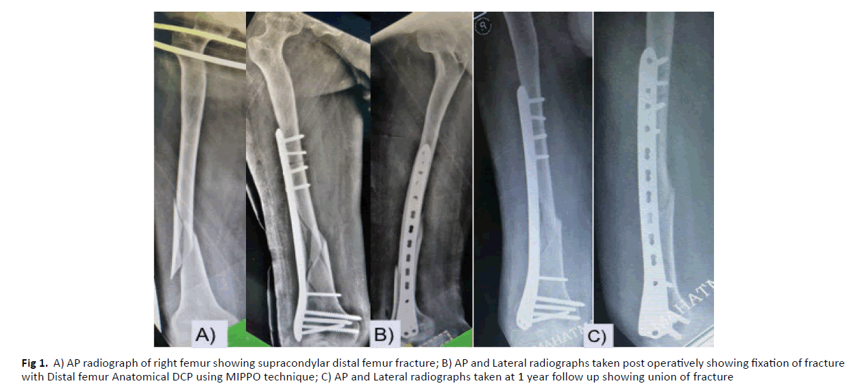
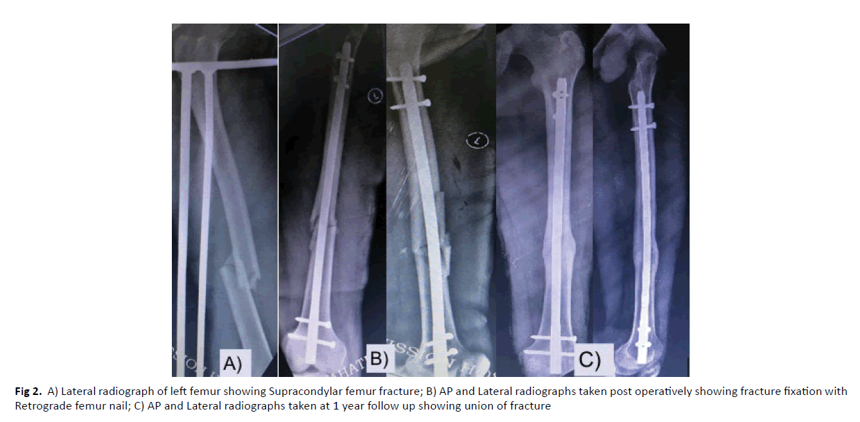
Numerous plating options have been proposed in the literature namely fixed and variable angled blade plates. However, these plates have been associated with higher rates of malunion, non-union implant failure, and infection owing to the rigid nature and associated soft tissue damage [19]. With the advent of minimally invasive plate osteosynthesis techniques, there has been a reduction in the aforementioned complications. These plates rely on the internal fixator principle and the fracture unites by callus formation because of the relative stabilization achieved with this fixation. (Figures 1 and 2)
Figure 1: A) AP radiograph of right femur showing supracondylar distal femur fracture; B) AP and Lateral radiographs taken post operatively showing fixation of fracture with Distal femur Anatomical DCP using MIPPO technique; C) AP and Lateral radiographs taken at 1 year follow up showing union of fracture
Retrograde intramedullary nailing being a load-sharing device has the advantage of bypassing the fracture site and providing a minimally invasive nature. However, they have an associated complication of knee arthrosis, stiffens, and mal-reduction.
In the present study, the prevalence of distal femur fractures was more in females, accounting for 50.70% of cases and there was a bimodal pattern involving more young adults and elderly females. Similar were the observations in previous studies [1,2,20]. The 33A2 fractures were more commonly observed in the present study involving more than half of the patient population. Similar findings were seen in the previous studies [5,8,20].
Previous studies have stated more than plating procedures have more blood loss as compared to nailing [7,15,21]. The observation can be related to the extensive soft tissue dissection which is commonly performed especially with the rigid plates. However, with the advent of less invasive stabilization techniques using locking plates, these issues have decreased substantially. A meta-analysis by Wang et al. [22] concluded that the intra-operative blood loss is less in patients treated by less invasive plating. Moreover, patients operated by retrograde nailing had more blood loss which was statistically significant in their study. The rate of blood loss in the present study was marginally more in patients with group A in present study, however, it was not statistically significant (p=0.8850). Apart from the blood loss, the intra-operative surgical timing also affects the outcome. The use of the aiming device with the plate and minimal soft tissue dissection can help reducing the surgical time as well as intra-operative blood loss. The operative duration between both the procedures has not been statistically significant in the previous studies [16,17,21,22]. On the contrary, patients in group A required longer surgical time, as opposed to patients in group B, which was statistically significant (p=0.0044). The meticulous dissection to avoid nail entry related complications increased fluoroscopy use for proper entry could be the potential reasons for the same.
Infection can be a threatening complication in any surgery. Nonetheless, because of the minimal soft tissue dissection and indirect reduction techniques, there has been a vast reduction in the same in the present scenario. There were 3 (4.22%) cases in group A and 1 (1.40%) cases in group B who had a superficial infection which was managed with oral antibiotics. There was no statistically significant difference in terms of infection as per the technique used. Similar findings have been reported in the literature previously [17,21,22]. There was no case of deep infection in either of the groups in the present study. Postoperative knee stiffness is a known complication associated with any open surgery around the knee joint. It can be postulated that extensive dissection can be a cause for the same. There were 6 (8.4%) cases in the present study that had post-operative stiffness and were left with 5 degrees and 10 degrees of extension lag. However, they were able to perform activities of daily living and had good signs of union and thus no further intervention was required. Meta-analysis and few studies have observed no statistically significant difference in terms of the technique used [17,11,22].
Fracture union remains to be one of the major concerns in distal femur fractures. Less stable fixations, increased soft tissue dissection is one of the major reasons for the delayed union, malunion, or non-union. The rate of non-union in patients treated locking plates varies between 1.6% and 6.1% [4,5,23]. Two systematic reviews [4,22], concluded that the rate of non-union was more in patients who were treated by locking plate which was as high as 5.3% as opposed to 1.5% in patients operated using intramedullary nailing. However, the difference was not statistically significant amongst both groups. Corollary to the above observation, the union in group A occurred early in the present study as compared to group B. Gill et al. [24] in their study comparing both the techniques have similar findings. They postulated that release of marrow contents at the fracture site and increased working length of the nail are factors that might lead to early union. Although the union rates in group A were marginally better in the present study, there was no statistically significant difference amongst both the groups. We did not have any case of delayed union, malunion, or non-union in the present study.
About 89% of the cases in the present study had excellent and good results as per the Schatzker and Lambert criteria. Nonetheless, there was no major difference between the groups in terms of the outcome.
This study is not without limitations. The small sample size and shorter duration of follow-up remain the limitations of the present study. More randomized trials and systematic reviews would help further to come to a definitive consensus.
Conclusion
Although intramedullary nailing is associated with the shorter operative time which can be statistically significant like in the present study, the final results of both nailing and plate osteosynthesis are similar with no implant superior to another. Both the techniques have similar complication rates.
REFERENCES
- Court-Brown C.M., Caesar B.: Epidemiology of adult fractures: A review. Injury.2006;37:691-697
- Martinet O., Cordey J., Harder Y., et al.: The epidemiology of fractures of the distal femur. Injury 2000;31:C62-C63
- Wahnert D., Hoffmeier K., Frober R., et al.: Distal femur fractures of the elderly-different treatment options in a biomechanical comparison. Injury. 2011;42:655-659
- Herrera D.A., Kregor P.J., Cole P.A., et al.: Treatment of acute distal femur fractures above a total knee arthroplasty: Systematic review of 415 cases (1981-2006). Acta Orthop. 2008;79:22-27
- Hierholzer C., von Rüden C., Pötzel T., et al.: Outcome analysis of retrograde nailing and less invasive stabilization system in distal femoral fractures: A retrospective analysis. Indian J Orthop. 2011;45:243-250
- Lupescu O., Nagea M., Patru C., et al.: Treatment options for distal femoral fractures. Maedica J Clin Med. 2015;10:117-122.
- Hartin N.L, Harris I., Kaushik H.: Retrograde nailing versus fixed angled blade plating for supra-condylar femoral fractures: A randomized controlled trial. ANZ J Surg.2006 ;76:290-294
- Gurkan V., Orhun H., Doganay M.,et al.: Retrograde intramedullary interlocking nailing in fractures of the distal femur. Acta Orthop Traumatol Turc. 2009;43:199-205
- Ostrum R.F, Maurer J.P.: Distal third femur fractures treated with retrograde femoral nailing and blocking screws. J Orthop Trauma. 2009;23:681-684
- Kolb W., Guhlmann H., Windisch C., et al.: Fixation of distal femoral fractures with the Less Invasive Stabilization System: A minimally invasive treatment with locked fixed-angle screws. J Trauma. 2008;65:1425-1434
- Smith T.O, Hedges C., MacNair R., et al.: The clinical and radiological outcomes of the LISS plate for distal femoral fractures: A systematic review. Injury. 2009;40:1049-1063
- Oh J.K., Hwang J.H., Sahu D., et al.: Complications rate and pitfalls of temporary bridging external fixator in periarticular comminutes fractures. Clin Orthop Surg. 2001;3:62-68
- Parekh A.A., Smith W.R., Silva S., et al.: Treatment of distal femur and proximal tibia fractures with external fixation followed by planned conversion to internal fixation. J Trauma. 2008;64:736-739
- Higgins T.F., Pittman G., Hines J., et al.: Biomechanical analysis of distal femur fracture fixation: fixed angle screw plate construct versus condylar blade plate. J Orthop Trauma. 2007;21:43-46
- Vllier H.A., Immler W.: Comparison of 95 degree angled blade plate and locking condylar plate for the treatment of distal femoral fractures. J Orthop Trauma.2012;26:327-332
- Markmiller M., Konrad G., Südkamp N.: Femur-LISS and distal femoral nail for fixation of distal femoral fractures: Are there differences in outcome and complications? Clin Orthop Relat Res. 2004;426:252-257
- Gao K., Gao W., Huang J., et al.: Retrograde nailing versus locked plating of extra-articular distal femoral fractures: Comparison of 36 cases. Med Princ Pract. 2013;22:161-166
- Greiwe R.M., Archdeacon M.T.: Locking plate technology: Current concepts. J Knee Surg. 2007;20:50-55
- Elsoe R., Ceccotti A.A., Larsen P.: Population-based epidemiology and incidence of distal femur fractures. Int Orthop. Jan. 2018;42:191-196
- Khursheed O., Wani M., Rashid S., et al.: Results of treatment of distal extra: Articular femur fractures with locking plates using minimally invasive approach-experience with 25 consecutive geriatric patients. Musculoskel Surg. 2014;99:139-147
- Rossas C., Nikolopoulos D., Liarokapis S., et al.: Retrograde intramedullary nailing vs. plat-ing in treatment of extrarticular distal femoral fractures: A comparative study. Injury. 2011;42:S20-S21
- Aijun W., Shuangle Z., Lixin S.,et al.: Meta-analysis of postoperative complications in distal femoral fractures: Retrograde intramedullary nailing versus plating. Int J Clin Exp Med. 2016;9:18900-18911
- Schandelmaier P., Partenheimer A., Koenemann B., et al.: Distal femoral fractures and LISS stabilisation. Injury. 2011;32:55-63
- Gill S., Mittal A., Raj M., et al.: Extra articular supracondylar femur fractures managed with locked distal femoral plate or supracondylar nailing: a comparative outcome study. J Clin Diagn Res. 2017.;11:RC19-RC23


 Journal of Orthopaedics Trauma Surgery and Related Research a publication of Polish Society, is a peer-reviewed online journal with quaterly print on demand compilation of issues published.