Hibernoma: unusual localization of a rare tumor
2 Department of Pathology, Fattouma Bourguiba University Hospital, Monastir, Tunisia, Email: abc@gmail.com
3 Department of Radiology, Lucie and Raymond Aubrac Hospital, Paris, France, Email: abc@gmail.com
Received: 18-Mar-2021 Accepted Date: Apr 26, 2021 ; Published: 05-May-2021, DOI: 10.37532/1897-2276.2021.16(1).16
This open-access article is distributed under the terms of the Creative Commons Attribution Non-Commercial License (CC BY-NC) (http://creativecommons.org/licenses/by-nc/4.0/), which permits reuse, distribution and reproduction of the article, provided that the original work is properly cited and the reuse is restricted to noncommercial purposes. For commercial reuse, contact reprints@pulsus.com
Abstract
Adipose tumors are dominated by lipomas, but other rarer entities may be encountered such as hibernoma which is a benign tumor that develops from brown fat. The thigh is the preferred location for hibernomas, but the tumor can appear in other areas. The upper limb is a rare localization and there is only one case of hibernoma on the hand already reported in the English literature. We report the case of a hibernoma of the palmar aspect of the hand. The tumor had no clinical specificities. MRI showed an uncertain appearance mimicking liposarcoma. The diagnosis was only confirmed by histological study. The treatment consisted of complete excision of the tumor. At a follow-up of 8 months, there was no recurrence.
Keywords
hibernoma, tumor, hand, treatment, surgery
Introduction
Hibernoma is a rare benign tumor of brown fat. The name originates from the similar appearance of these tumors to hibernating glands in animals, which appear light brown and lobulated. Since the first description by Merkel in 1906, less than 250 cases have been reported in the English literature. The tumor is often described as an asymptomatic mass of soft tissue, located in a proximal position, where the brown fat persists, thus the thigh is the preferential location for hibernomas. The localization in the hand is extremely rare and to our knowledge, just one case of a dorsal hibernoma of the thumb has been documented.
Clinically, the tumor has no specific symptoms. In the MRI, differential diagnoses include lipoma and liposarcoma. The diagnosis is confirmed by a histological study. Complete surgical excision is the mainstay of treatment.
We report a case of Hibernoma of the thenar eminence.
Case Report
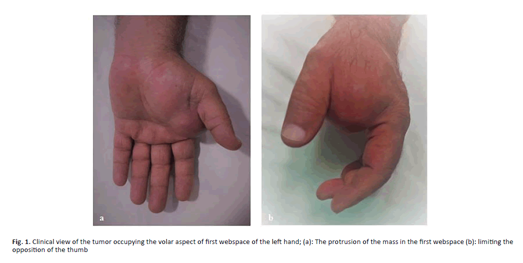
The patient was a 61-year-old, without medical history, complaining of a subcutaneous tumor filling the palmar aspect of the first web space of the left hand. The painless lump had appeared five years ago and gradually increased in size. The skin above was tight with normal coloration. The tumor was soft and fixed (Figure 1a).
The protrusion of the mass which measured 7 cm × 4 cm was limiting the opposition of the thumb. There was no neurological complaint (Figure 1b).
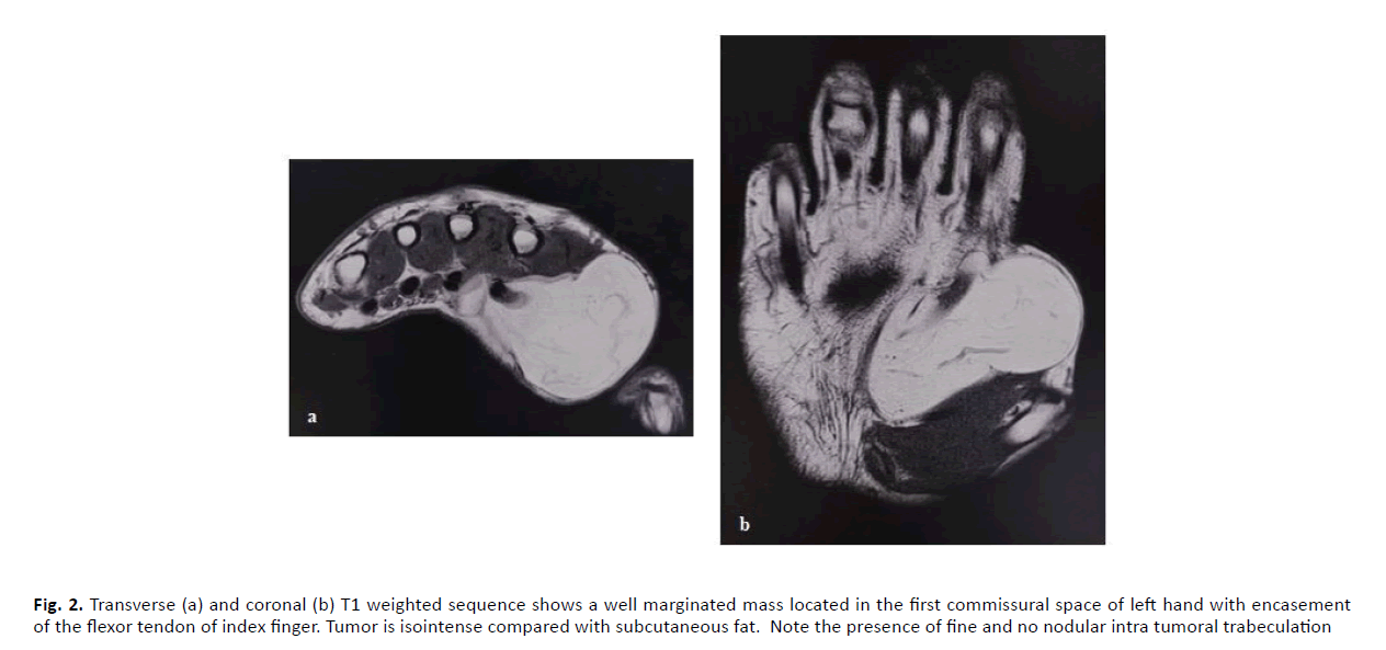
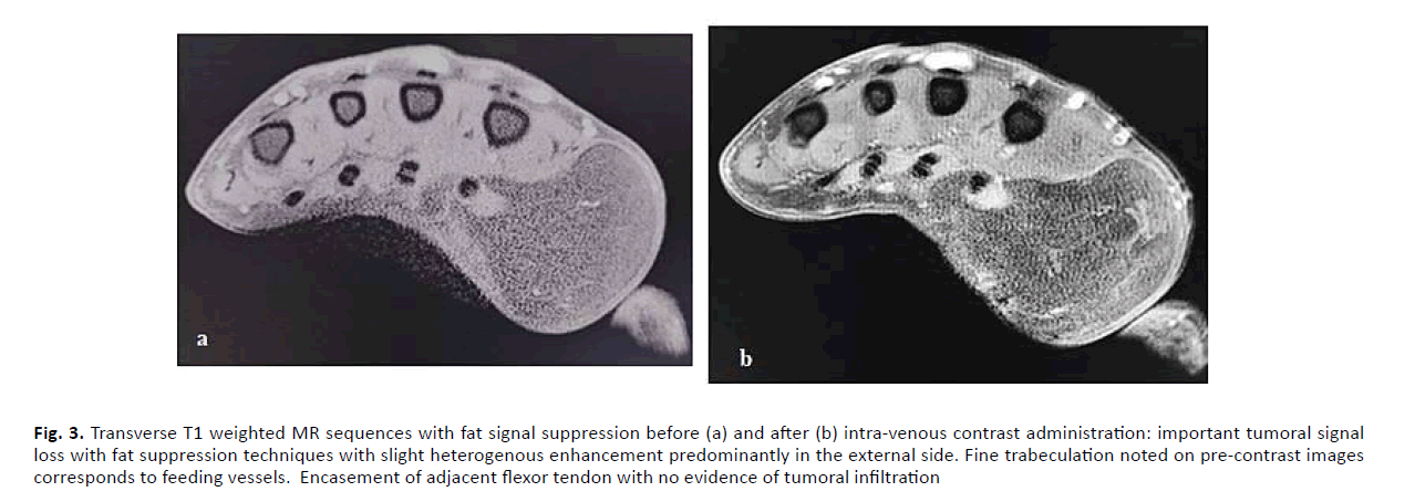
The MRI showed a subcutaneous limited fat-containing mass which measured 80 mm × 35 mm, crossed by vascular structures (Figures 2 and 3). The tumor reached the carpal tunnel and surrounded the volar aspect of the median nerve and the flexor tendons of the second, third and fourth finger. The main differential diagnosis was liposarcoma on account of the size and the vascularization.
Figure 2: Transverse (a) and coronal (b) T1 weighted sequence shows a well marginated mass located in the first commissural space of left hand with encasement of the flexor tendon of index finger. Tumor is isointense compared with subcutaneous fat. Note the presence of fine and no nodular intra tumoral trabeculation
Figure 3: Transverse T1 weighted MR sequences with fat signal suppression before (a) and after (b) intra-venous contrast administration: important tumoral signal loss with fat suppression techniques with slight heterogenous enhancement predominantly in the external side. Fine trabeculation noted on pre-contrast images corresponds to feeding vessels. Encasement of adjacent flexor tendon with no evidence of tumoral infiltration
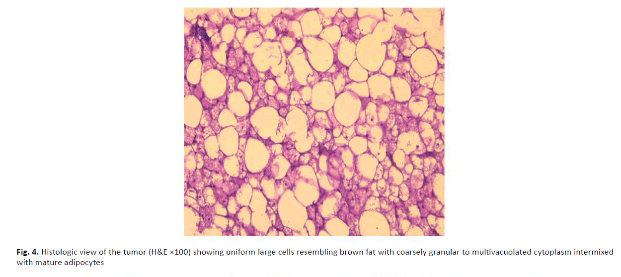
We proceeded by a surgical biopsy. In histological examination, the tumor contained uniform large cells resembling brown fat intermixed with mature adipocytes.
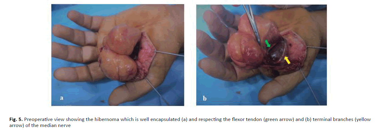
Two weeks later, the patient underwent resection of the mass under regional anesthesia. A 7 cm volar incision was required. The tumor was easily dissected and completely excised despite its close relationship with the flexor tendon and the median nerve (Figure 4). Feeding vessels were ligated. After excision of the excess skin, the incision was closed. In histological examination, the mass was encapsulated and lobulated formed by brown fat and some mature adipocytes separated by fibrous septa, including vessels and inflammatory cells. There is no atypia. (Figure 5).
There was no postoperative complication, and the patient retrieved a complete opposition of the thumb in few days. At an 8-month-followup, there is no recurrence.
Discussion
Hibernomas are benign soft-tissue tumors emerging from vestigial brown fat, which can be located in the subcutaneous tissue, the skeletal muscle, or the intermuscular fascia [1].
This tumor is uncommon and accounts for <2% of all adipocytic tumors [2].
Brown fat is crucial in non-shivering thermogenesis in hibernating animals and the human fetus [3].
The high concentration of mitochondria and hypervascularization leads to the dark color, which differentiates it from white adipose tissue [4].
The largest and most valid case series of hibernomas was 170 patients by Furlong MA et al. [1], who proved that the common location is the thigh, followed by upper extremity, trunk, neck, then abdominal cavity or retroperitoneum.
These tumors affect mainly adults in the 3rd and 4th decades of life with a slight male predominance [1,2,5].
The clinical presentation may vary depending on the location. The tumor is often asymptomatic and present as soft, painless, palpable, and mobile masses. They become symptomatic only when associated with compression of surrounding structures due to their growth [1,4,6].
Superficial hibernomas can be considered as lipomas, which are the same in presentation and much more common [4].
The diagnosis of hibernoma is difficult. Radiographs may show a radiolucent mass, typically without calcification or bone erosion [7]. The ultrasound exam shows a consistently hyperechoic mass with hypervascularization and enlarged vessels [8].
The MRI is the essential morphologic investigation, allowing a better characterization of the size and location of the tumor, and establishing the differential diagnosis with malignant tumors.
Hibernomas are slightly or distinctly hypointense to subcutaneous fat on T1W images and don’t fully suppress on STIR or fat-saturated T2W images [8,9].
The differential diagnosis of a complex lipomatous tumor on MRI is wide and involves benign conditions, such as lipoma, angiolipoma, hemangioma, and malignant tumors such as liposarcoma.
Poorly differentiated (high-grade) liposarcomas are rarely confused with lipomas, or hibernomas, as they include slight or no fatty components.
Otherwise, the fatty component of a well-differentiated liposarcoma appears isointense to subcutaneous fat, on T1WSE, distinguishing them from hibernomas [8].
The definitive diagnosis of Hibernoma is based on pathology. Grossly, the tumor is well-encapsulated, containing white and brown fat separated by fibrous septa and vessels. Its color may range from yellow to brown. On the microscopic examination, there are three principal cell types: cells with granular eosinophilic cytoplasm containing few lipid vacuoles; larger cells with multiples vacuoles; and univacuolated large cells. Generally, there is no mitosis or atypia [4].
Histologically, the main differential diagnosis is the atypical lipomatous tumor, which is eliminated by negative MDM2 and CDK4 on immunohistochemistry [5].
The surgical treatment consists of a monobloc excision of the tumor which must be ideally done with minimal dissection. The resected hibernoma is usually a smooth, well-encapsulated, and non-adherent mass.
This excision is curative and has a low risk for local recurrence. When the diagnosis is uncertain, accurate hemostasis should be achieved and the patient should receive further evaluation and possibly treatment before definitive surgical management such as our case.
The diagnosis is confirmed by histopathological examination of the resected tumor.
Incomplete excision may occur in continued growth and recurrence, even in tumors that appear to be histologically benign.
In all published reports of hibernomas in humans in the English literature, no cases have been reported of malignant transformation or distant metastasis.
However, two cases of recurrence are reported, because of incomplete excision [10].
Conclusion
Hibernoma is communally located in axial position, but it can exist elsewhere. The hand is an exceptional location of the tumor. Hibernoma has no clinical specificities and can be considered as lipoma. In our case, the tumor was in close relation with the median nerve; however, there is no neurological complaint. The MRI confirms the lipomatous nature of the tumor but it cannot decide between hibernoma and other diagnoses such as liposarcoma. Only histological study can confirm the diagnosis. Surgical monobloc resection is the main treatment and recurrence depends on the quality of the excision.
REFERENCES
- Furlong M.A., Fanburg-Smith J.C., Miettinen M.: The morphologic spectrum of hibernoma: a clinicopathologic study of 170 cases. Amer J Sur Pathol. 2001;25:809-814.
- Fletcher C.D., Unni K.K., Mertens F.: Pathology and genetics of tumours of soft tissue and bone. IARC; 2002.
- Dardick I.: Hibernoma: A possible model of brown fat histogenesis. Human Pathol. 1978;9:321-329
- Cipriano C.A., Gray R.R., Fernandez J.J.: Hibernomas of the upper extremity: A case report and literature review. Hand. 2015;10:547-549.
- Al Hmada Y., Schaefer I.M., Fletcher C.D.: Hibernoma mimicking atypical lipomatous tumor: 64 cases of a morphologically distinct subset. Amer J Surg Pathol. 2018;42:951
- Greenbaum A., Coffman B., Rajput A.: Hibernoma: diagnostic and surgical considerations of a rare benign tumour. Case Reports. 2016;2016:bcr2016217625
- Kransdorf M.J., Bancroft L.W., Peterson J.J., et al.: Imaging of fatty tumors: distinction of lipoma and well-differentiated liposarcoma. Radiol. 2002;224:99-104
- Lee J.C., Gupta A., Saifuddin A., et al.: Hibernoma: MRI features in eight consecutive cases. Clinical Radiol. 2006;61:1029-1034
- Liu W., Bui M.M., Cheong D., et al.: Hibernoma: comparing imaging appearance with more commonly encountered benign or low-grade lipomatous neoplasms. Skeletal Radiol. 2013;42:1073-1078
- Beals C., Rogers A., Wakely P., et al.: Hibernomas: a single-institution experience and review of literature. Med Oncol. 2014;31:769



 Journal of Orthopaedics Trauma Surgery and Related Research a publication of Polish Society, is a peer-reviewed online journal with quaterly print on demand compilation of issues published.