Reliability of the ultrasound measurements of deep abdominal muscle in rehabilitative practice
- *Corresponding Author:
- Dr. Katarzyna Szušcik
Department of Adapted Physical Activity and Sport, School of Health Sciences in Katowice, Medical University of Silesia in Katowice, Poland
Tel: +48 695182050
E-mail: kszuscik@sum.edu.pl
Received Date: 14.03.2017 Accepted Date: 27.04.2017 Published Date: 29.04.2017
Abstract
Introduction: Deep abdominal muscles play an important role in controlling the lumbosacral and hip regions and maintaining core stability. As they take part in the muscle bracing, their activation and efficiency increase are elements of prevention and therapy, e.g. in spine-related painful conditions. Complementing traditional therapy with the ultrasound muscle assessment applied both in the course of therapy and as an element of evaluation of its findings may improve the effectiveness of physiotherapeutic procedures. Research objective: The objective of the study was to assess the reliability of measuring of the lateral abdominal wall deep muscles by means of an ultrasound machine in accordance with the modified research procedure. Material and methods: The study comprised 90 people, where 20 (13 women and 7 men) were selected in random to be included in statistical analysis. Their results were used as the basis for calculating the intraclass correlation coefficient ICC 3,1. Apart from a range of preliminary tests including taking the body weight, WHR ratio and skinfold measurement, every person subject to the study underwent an assessment of transverse sections of their lateral abdominal wall deep muscles (TVA, IO, EO, and their common measurement). The test was always conducted by one, the same researcher. Each measurement was taken 5 times with vertical placing transducer and conscientiously following the research procedure presented in the article. Results: Descriptive statistics have been prepared. The average age of the subjects was around 23.5 years (SD 3.14). The reliability of measurements obtained in the studied population proved to be very high and amounted to ICC=0.98. Conclusion: It was achieved very high reliability of measurements in a modified test procedure. Repeating the measurements of deep abdominal muscles five times and with vertical placing transducer may still increase the reliability of the research findings. In view of this high reliability, ultrasound appears to be an objective and available method enriching diagnostics and treatment of muscle dysfunctions in rehabilitative practice.
Keywords
Ultrasound, Abdominal muscles, Reliability
Introduction
Application of Rehabilitative Ultrasound Imaging (RUSI), both in practice and within scientific research, is evidence of the continuous development of physiotherapy [1]. Progress may also be observed in ultrasonography; it is worth noting that in 2007, when Teyhen et al. published some of the first articles on the ultrasound assessment of abdominal muscles, it was a convex transducer that was used in the examinations [2]. At present, in order to evaluate the musculoskeletal system, and thus the tissues and structures located superficially, it is recommended to apply higher frequency linear transducers [3-5]. It needs to be emphasized that an ultrasound examination enables therapists to assess the structures in motion; as a functional test, it is mainly appreciated in the musculoskeletal system functional diagnostics [1,4,6,7].
The first articles devoted to the topic of ultrasound assessment of the muscles came out at the end of the 1990s (Hodges, Whittaker, Ferreira) [2,8,9]. Their findings contributed to broadening the knowledge of functional significance of abdominal muscles in controlling the lumbosacral and hip regions and maintaining core stability. Complex work on strengthening, activating and enhancing efficiency of the muscles taking part in the so-called muscle bracing is recommended in prevention of spine-related painful conditions and in rehabilitating individuals after injuries of the locomotor system [10,11]. Moreover, ultrasound is a renowned method for activating deep abdominal muscles which is focused on both the quantitative assessment (by measuring the muscle cross sections) and the qualitative evaluation (by determining the character of muscle fibre activation). Qualitative assessment is also a valuable addition to the course of therapy using the sonofeedback method [1,12,13].
The objective of the study was to assess the reliability of measuring thickness of the lateral abdominal wall deep muscles by means of an ultrasound machine in accordance with the modified research procedure with repeating the measurements five times and with vertical placing transducer. The article describes in detail the methodology of measuring the thickness of the abdominal muscles using ultrasound to increase the level of knowledge and practical skills of physiotherapists in their daily practice. In chapter discussion were described procedures already known, previously used and published in the literature.
Material and Methods
Ninety out of 102 people that enrolled as participants of the study (60 women and 30 men), underwent the complete research procedure. Inclusion criteria comprised the following: the age of 18-45 years, the absence of a recognized chronic disease capable of affecting the course and results of the study, and the absence of spine-related pain or the occurrence of pain without associated neurological symptoms. Exclusion criteria included the age ≤18 and ≥45, severity of the lumbar spine pain ≥7 points in VAS (Visual Analog Scale) at the moment of the procedure, presence of spinerelated painful conditions with associated neurological symptoms, the occurrence of a chronic disease able to affect the course and result of the study, past surgical procedures within the abdominal cavity, the skinfold measurement <30 mm and pregnancy in the females.
In order to assess the reliability of measurements, 20 people were selected at random, and their results were used in statistical analysis.
The study participants were recruited by means of announcements about the research project in the form of posters presented in the university campus area.
The research project was accepted by the Bioethics Committee of the Medical University of Silesia in Katowice (KNW/0022/KB46-7/15).
Equipment
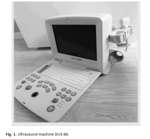
In the study we used a portable ultrasound machine Edan DUS 60 with a linear transducer of the frequency range of 6-10 MHz, working in the B-mode (Figure. 1). The transducer was 40 mm long. The measurements were taken with the machine accuracy of 0.01 mm, and with this accuracy noted down in research questionnaires by the researcher. The frequency of the transducer work amounted to 10MHz, and the depth of ultrasound wave’s penetration was set at up to 39 mm or 49 mm, depending on the thickness of a studied person’s tissues. Additionally, we introduced slight modifications in the settings of the machine, among others, in the shades of grey or the level of focusing, all in order to obtain the best possible quality of the image.
Researcher
The examinations of transverse sections of deep abdominal muscles were all taken by the same researcher, who has been performing ultrasound examinations of the musculoskeletal system for 18 months, and has completed the training in ultrasound imaging of the musculoskeletal system with elements of rheumatology conducted by the Polish Ultrasound Society.
Research Procedure
Before starting the proper study, a team of researchers conducted a pilot study lasting for 4 weeks and comprising 5 people chosen at random, in order to work out measurement procedures and identify potential errors.
The procedures were carried out in a diagnostic room of the Medical University of Silesia, in the same conditions and with the same standards for every participant.
The study participants got familiar with the methodology and provided with a short study description. After signing a written consent concerning participation in the project, each person was interviewed. The questions concerned, among others, sociodemographic data, the occurrence and severity of spinerelated painful conditions and other diseases, past injuries and surgical operations. After preliminary qualifications one of the researchers performed the chosen measurements.
Following taking the height by means of a height measure, the body weight and composition were assessed with the use of a Tanita®- BC-418 analyser.
Next a metric tape measure was applied to take the waist and hip circumferences, with regard to the following anatomical points: for the waist circumference it was the level of the waist indentation; to take the hip circumference the tape measure was placed at the level of the greater trochanters of the femurs. The measurements were taken in a relaxed standing position with the feet hip-width apart. To avoid a measurement error, each measurement was repeated three times.
Bearing in mind the influence of the adipose tissue on deteriorating the quality of ultrasound imaging, in the research methodology we also took into consideration measuring the adipose tissue distribution at the site of the future ultrasound transducer application. The skinfold measurement was performed by means of an analogue skinfold caliper. Using both hands, one researcher grabbed a skinfold above the right anterior superior iliac spine and at the level of the umbilicus, while another researcher applied the caliper and read the value. This measurement was also performed three times in the abovementioned body position.
The results of all the measurements were noted down in the study questionnaire. The next stage of the study was the ultrasound analysis of deep muscles of the lateral abdominal wall. The manner in which the measurements were taken and a choice of the examination body position were based on a profound analysis of research results published by Hodges, Whittaker, Ferreira, Costa, Gnat, and Saulicz, as well as the researchers’ own experience gained in physiotherapeutic practice and the pilot study [1,2,3,14]. Measurement methodology was modified with a new vertical placing transducer and the number of measurements.
The assessment concerned thickness of abdominal muscles, including the transversus abdominis (TVA), internal oblique (IO) and external oblique muscles (EO).
Each examined person assumed the supine position, lying on their back on a couch, with their upper extremities at their sides, and with the anterior abdomen exposed from the costal margin to the anterior superior iliac spines.
The cervical spine of the examined person was supported with a cushion which neutrally filled the cervical lordosis for increasing comfort during the examination. The knee joints were supported with a 20-cm-high semiroller ensuring the knee flexion of around 15-30 degrees, which allowed for relaxation of the anterior and posterior musculofascial trains. In every person, the same cushion and roller were used. During the examination, the subjects were asked to relax, breathe freely and remain in the position throughout the procedure.
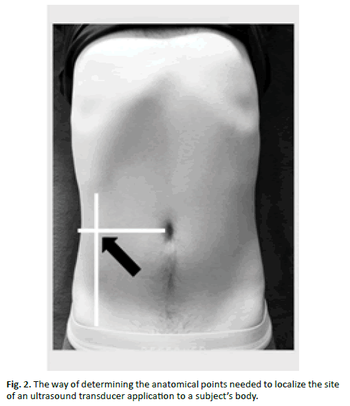
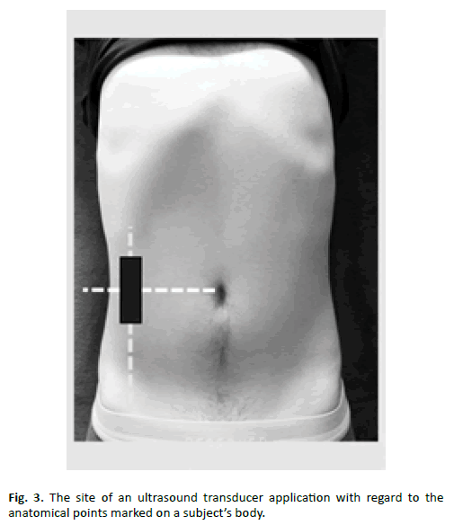
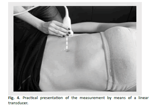
The site of application of the transducer is presented in Figures. 2-4. The measurement was always performed on the right side of the body - the assumption of symmetry of the abdominal muscles on both sides of the body [15]. The measurement point was determined by palpating two topographic points within the abdomen: the umbilicus and the right anterior superior iliac spine. Starting with the umbilicus, the researcher determined the first line running sideways, perpendicularly to the midline of the body (which anatomically marks the sagittal body axis containing the umbilicus). The second line was determined starting with the anterior superior iliac spine and running upwards, parallelly to the midline of the body and perpendicularly to the first line. The point where the lines crossed at the angle of 90 degrees constituted the measurement point. The transducer was applied in a different way than described in literature - the vertically - in cross-section transversely to the fibres of the transversus abdominis.
The research procedure consisted in taking 5 independent measurement cycles (P1-P5), where one measurement followed another. In every cycle, after freezing the image, the researcher put the transducer aside, took the measurements, noted the results in the questionnaire, and proceeded to the next measurement cycle.
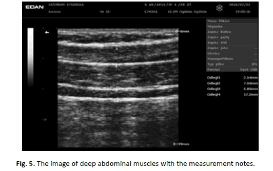
In every cycle the thickness of three muscles (TVA, IO, EO) was measured separately. The measurements did not include the intermuscular septa, which distinctly bordered the muscle bellies with hyperechogenic shadows. The researcher also performed measurements of all the three muscles together, from the inferior edge of the transversus abdominis to the superior edge of the external oblique muscle (TVA+IO+EO), including 2 intermuscular septa, between the transversus abdominis and internal oblique, and between the internal oblique and external oblique muscles (Figure. 5). Intermuscular septum are clearly visible as a hyperechoic streaks. This measure is justified by the inter-individual variability in the construction of intermuscular septum described in the article Urquhart’a, wherein between the transverse abdominal muscle and internal oblique muscles may be intermuscular septum division into two layers, which may affect the thickness of the entire complex deep abdominal muscles [16].
Taking into account the respiratory function of deep abdominal muscles, the researcher observed the subject’s chest and froze the image (pressed the „freeze” button) at the end of their exhalation, repeating this observation before every subsequent freezing of the image.
It needs to be emphasized that the researcher applied the transducer to the patient’s body without additional pressure, in his own idiosyncratic way, placing it as transversely to the examined structures as possible, and avoiding the phenomenon of anisotropy. The way of handling the transducer was a standard one, that is, with the fingers on the ulnar side, directly by the skin of the subject, and holding the transducer with the fingers on the radial side (between the thumb and index finger).
Statistical Analysis
The statistical analysis comprised 19.6% of all the results (20 people). The intraclass correlation coefficient (ICC) was calculated. We made use of model 3 (ICC 3,1), which is applied when one and the same researcher performs all the measurements on his or her own. Therefore, randomness concerns only the study findings and not a variety of several researchers. To carry out the statistical analysis Statistica 10.0 programme was used. The calculated values included arithmetic means, standard deviations, minimum and maximum values and the median [17,18].
Results
The descriptive statistics characterizing a group of randomly chosen people (13 women and 7 men) are summarised in Table 1. The average age of the studied subjects was 23.5 years (SD 3.14).
| Variables | Gender | N | Mean | Median | Min | Max | Standard deviation |
|---|---|---|---|---|---|---|---|
| Age [years] |
females | 13 | 22.77 | 22 | 21 | 30 | 3.03 |
| males | 7 | 24.86 | 26 | 20 | 28 | 3.08 | |
| Body mass [kg] |
females | 13 | 58.49 | 59.2 | 47.8 | 81.7 | 14.10 |
| males | 7 | 78,91 | 75.3 | 67.7 | 102 | 11.26 | |
| Body height [cm] |
females | 13 | 166.07 | 167 | 153 | 179 | 7.82 |
| males | 7 | 181.42 | 179 | 173 | 193 | 7.36 | |
| BMI [kg/m2] |
females | 13 | 22.44 | 22.6 | 19.4 | 26.1 | 2.51 |
| males | 7 | 23.99 | 23.5 | 18.7 | 28 | 2.89 | |
| Waist circumference [cm] |
females | 13 | 70.92 | 71 | 64 | 82 | 4.94 |
| males | 7 | 83.71 | 83 | 75 | 96 | 6.82 | |
| Hip circumference [cm] |
famales | 13 | 99.17 | 99 | 92 | 110 | 6.22 |
| males | 7 | 98.85 | 99 | 91 | 108 | 6.82 | |
| WHR ratio | females | 13 | 0.71 | 0.71 | 0.67 | 0.78 | 2.03 |
| males | 7 | 0.84 | 0.84 | 0.82 | 0.88 | 2.02 | |
| Skinfold thickness [mm] | females | 13 | 18.62 | 18 | 9 | 26 | 5.42 |
| males | 7 | 16.14 | 16 | 10 | 23 | 5.87 |
Table 1. Descriptive statistics of the subjects in view of gender.
In order to assess reliability of the measurements, that is, to determine the intraclass correlation coefficient (ICC), we used the results of the abdominal muscles transverse sections, which are presented in Table 2. Comparing to the group of women, in the group of men there are, on average, larger thickness of each muscle measured separately, as well as of all of them measured together. The thickest in the whole complex of the lateral abdominal wall muscles is the internal oblique muscle. In terms of its thickness, it constitutes 40.56% and 45.39% of the whole complex (TVA+IO+EO) in the women and men, respectively.
| Muscle | Gender | N | Mean | Median | Min | Max | Standard deviation |
|---|---|---|---|---|---|---|---|
| TVA transversus abdominis |
females | 13 | 3.01 | 3 | 1.54 | 4.03 | 0.77 |
| males | 7 | 3.14 | 3.38 | 2.21 | 3.84 | 0.58 | |
| IO internal oblique |
females | 13 | 8.51 | 8.45 | 5.69 | 11.4 | 1.32 |
| males | 7 | 11.15 | 10.8 | 7.46 | 14.9 | 2.37 | |
| EO external oblique |
females | 13 | 7.96 | 7.01 | 5.3 | 13.2 | 2.53 |
| males | 7 | 8.67 | 8.37 | 6.16 | 12.6 | 1.86 | |
| TVA+IO+EO common measurement |
females | 13 | 20.98 | 20 | 14.9 | 27.3 | 3.84 |
| males | 7 | 24.56 | 23.4 | 17.7 | 31.9 | 4.57 |
Table 2. Mean results of thickness of the abdominal muscles [mm].
The results regarding assessment of the intraclass correlation coefficient (ICC) are summarized in Tables 3 and 4. The findings are presented for 5 measurements of particular muscles, taking into account 20 cases and 1 researcher. Each measurement is highly reliable. For each measurement, the intraclass correlation coefficient is 0.99.
| Muscle | Mean result for ICC 3,1 |
|---|---|
| TVA transversus abdominis |
0.9917 |
| IO internal oblique |
0.9871 |
| EO external oblique |
0.9942 |
| TVA+IO+EO common measurement |
0.9947 |
Table 3. Results of the intra-class correlation coefficient (ICC 3,1) for 20 randomly selected results.
| Measure-ment number | N | Mean | Confidence interval -95.000% |
Confidence interval 95.000% |
Min | Max | SD | Confidence interval SD -95.000% |
Confidence interval SD 95.000% |
Standard error |
|---|---|---|---|---|---|---|---|---|---|---|
| TrA1 | 20 | 2,99250 | 2,66332 | 3,32168 | 1,54000 | 4,07000 | 0,703352 | 0,534892 | 1,027296 | 0,157274 |
| TrA2 | 20 | 3,02950 | 2,69449 | 3,36451 | 1,54000 | 4,07000 | 0,715810 | 0,544367 | 1,045492 | 0,160060 |
| TrA3 | 20 | 3,06800 | 2,72859 | 3,40741 | 1,63000 | 4,15000 | 0,725219 | 0,551522 | 1,059235 | 0,162164 |
| TrA4 | 20 | 3,08200 | 2,74335 | 3,42065 | 1,73000 | 4,15000 | 0,723592 | 0,550285 | 1,056858 | 0,161800 |
| TrA5 | 20 | 3,15000 | 2,80989 | 3,49011 | 1,83000 | 4,30000 | 0,726709 | 0,552655 | 1,061410 | 0,162497 |
| OI1 | 20 | 8,96700 | 7,95910 | 9,97490 | 5,69000 | 13,80000 | 2,153565 | 1,637766 | 3,145436 | 0,481552 |
| OI2 | 20 | 9,22600 | 8,21666 | 10,23534 | 5,76000 | 14,20000 | 2,156643 | 1,640106 | 3,149932 | 0,482240 |
| OI3 | 20 | 9,40450 | 8,41536 | 10,39364 | 5,84000 | 14,40000 | 2,113474 | 1,607277 | 3,086881 | 0,472587 |
| OI4 | 20 | 9,62700 | 8,58971 | 10,66429 | 5,99000 | 14,70000 | 2,216368 | 1,685526 | 3,237164 | 0,495595 |
| OI5 | 20 | 9,94050 | 8,89008 | 10,99092 | 5,99000 | 14,90000 | 2,244414 | 1,706855 | 3,278128 | 0,501866 |
| OE1 | 20 | 7,89250 | 6,81763 | 8,96737 | 5,30000 | 13,00000 | 2,296659 | 1,746587 | 3,354434 | 0,513548 |
| OE2 | 20 | 8,08550 | 6,99781 | 9,17319 | 5,45000 | 13,00000 | 2,324049 | 1,767417 | 3,394440 | 0,519673 |
| OE3 | 20 | 8,23400 | 7,13420 | 9,33380 | 5,53000 | 13,00000 | 2,349921 | 1,787093 | 3,432229 | 0,525458 |
| OE4 | 20 | 8,35400 | 7,22369 | 9,48431 | 5,53000 | 13,10000 | 2,415120 | 1,836675 | 3,527456 | 0,540037 |
| OE5 | 20 | 8,48000 | 7,33415 | 9,62585 | 5,45000 | 13,20000 | 2,448323 | 1,861926 | 3,575951 | 0,547462 |
| TRA+OI+OE1 | 20 | 21,58000 | 19,56101 | 23,59899 | 14,90000 | 30,20000 | 4,313943 | 3,280712 | 6,300823 | 0,964627 |
| TRA+OI+OE2 | 20 | 21,93500 | 19,88369 | 23,98631 | 15,00000 | 30,70000 | 4,383014 | 3,333240 | 6,401707 | 0,980072 |
| TRA+OI+OE3 | 20 | 22,24000 | 20,12460 | 24,35540 | 15,10000 | 31,50000 | 4,519944 | 3,437374 | 6,601702 | 1,010690 |
| TRA+OI+OE4 | 20 | 22,49000 | 20,33757 | 24,64243 | 15,20000 | 31,70000 | 4,599073 | 3,497551 | 6,717276 | 1,028384 |
| TRA+OI+OE5 | 20 | 22,90500 | 20,71921 | 25,09079 | 15,30000 | 31,90000 | 4,670340 | 3,551749 | 6,821367 | 1,044320 |
Table 4. Confidence interval.
Discussion
When analysing the measurement values of deep abdominal muscles transverse sections, it is worth paying attention to their very high reliability obtained both in the present study and in the mentioned studies by other authors, what seems to be the result of modified test procedure.
There are 3 paths in the area of research concerning evaluation of reliability and compatibility of deep muscle ultrasound measurements that attract attention while reviewing the literature: 1. The assessment of muscle thickness in contraction and release phases in static positions, 2. The assessment of changes in the muscle thickness with regard to differences between their tension and relaxation conditions, 3. The assessment of changes of the muscle thickness over a longer period of time [19].
In the study of Costa et al. the ICC 2.1 coefficient was calculated based on the evaluation of thickness of deep abdominal muscles in a static image and supine position, and amounted to 0.97. The cited study also assessed the reliability of measurements taken in progress of changes in the muscle thickness (in the phases of contraction and release), where ICC was 0.72, which implies lower reliability of the measurement [10]. In another article by Mangum et al. 16 healthy people were subject to study. The intraclass correlation coefficient (ICC 3, k) was determined for measuring the thickness of the transversus abdominis and multifidus muscles. For TVA the obtained reliability was nearly perfect, at the level of 0.90. The measurement was also taken in a supine position, where the reliability of measurements proved to be higher than in other positions (in a sitting position ICC3, k=0.61, in standing ICC3, k=0.55, and while walking on a treadmill ICC3, k=0.74). Interestingly, while walking on a treadmill, the obtained measurement reliability was higher than in static sitting and standing positions. For the multifidus muscle, the value of the ICC coefficient was only 0.26, which implies a very low reliability thereof. This value concerns the measurement taken in a supine lying position; in other positions the ICC result was even lower [20].
In another study authors Gnat et al. characterized the two researchers - fully-qualified physiotherapists, who nevertheless had very limited experience in performing ultrasound examinations and interpreting them. The researchers completed two weeks’ training supervised by a specialist in ultrasonography. One of the analysed results was the ICC coefficient for the measurement of the TVA thickness, who showed high reliability study [21]. In the present study, every person underwent 5 measurements one after another, which gives a choice between 3 and 5 measurement or calculation of average values at the stage of data analysis and might have contributed to obtaining very high reliability of the study results. Moreover, at the stage of data acquisition can exclude the measurement error occurs by the investigator.
The so far published studies have also varied in research procedures, depending on the patients’ starting positions in which the muscle ultrasound assessment was performed, and the methods of determining the measurement site.
An interesting subject’s body position was suggested by Ferreira et al. [22]. He described a supine lying position with the upper limbs crossed over the chest and the lower limbs flexed at the angles of 50 degrees in hips and 90 degrees in knees, additionally suspended with a system of ropes. This position enabled the patient to relax the trunk and lower extremities and simultaneously perform movement tasks also evaluated in the study, and reduced friction. During the examination researchers determined a line connecting the costal margin and the wing of the ilium, and another one running laterally to the midline of the body. A transducer was applied 10cm from the body midline, transversely to the muscle fibres. The researchers used a convex transducer. A similar method of determining the measurement site was described in the study by Gnat at al., where one line was marked running sideways from the umbilicus, and the other – running cranially from the anterior superior iliac spine on the right side, and where the measurement site was a crossing point between the two [21]. A similar methodology of determining the measurement site was used in the present study. In the article by Linek et al. [23], which describes the assessment of adolescents aged 10 to 16, the site of measuring deep abdominal muscles was also determined on the anterolateral abdominal wall, between the wing of the ilium and the costal margin, parallel to the longitudinal body axis. The assessment of the muscle thickness was performed in a supine lying position, with the upper limbs at the sides, and with the lower limbs extended both in hips and knees, which is different from the other cited studies.
The argument for a new vertical placing transducer, was found in a study Urquhart et al., which rated on cadavers morphology of the deep abdominal muscles including the direction of the fascia [16]. It has been shown that fascia of TrA has a horizontal course most marked in the central region of the lateral abdominal wall (region set between the lower limit of the last rib and the anterior superior iliac spine) - where the measurements were taken. According to the methodology ultrasound muscular system it is recommended to take measurements using mainly cross-section relative to the muscle fibers [24]. To confirm the validity of this thesis seems to be reasonable to compare in the future the results obtained, both in setting the horizontal and vertical placing transducer in one study.
Summarizing our own study results, as well as the findings of the cited researchers, it is possible to claim that ultrasound constitutes a reliable measuring tool, which perfectly complements the muscle assessment, especially in the case of the lateral abdominal wall muscles. The assessment of the deep abdominal muscles thickness appears to be a method available to all researchers, including those who have only completed a training of ultrasound imaging of body structures, and who use their skills just within the framework of scientific research.
Conclusion
1. Ultrasound assessment of transverse sections of the transversus abdominis, internal oblique and external oblique muscles reveals high measurement reliability.
2. Five-fold measurement of thickness of deep abdominal muscles muscles can be used as an alternative measurement of three-fold in order to achieve even greater reliability testing.
3. The vertical position of probe seems to be an alternative method of measuring the thickness of the side wall of the abdominal muscles indicating a very high reliability of measurements.
4. Due to high reliability of measuring deep abdominal muscles, ultrasound seems to be an objective and available method which complements diagnostics and treatment of muscle dysfunctions in physiotherapy and rehabilitation.
References
- Ferreira P., Ferreira M., Nascimento D., et al.: Discriminative and reliability analyses of ultrasound measurement of abdominal muscles recruitment. Man Ther. 2011 16: 463-469.
- Teyhen D., Gill N., Whittaker J., et al.: Rehabilitative ultrasound imaging of the abdominal muscles. J Orthop Sports Phys Ther. 2007 37: 450-466.
- Costa LOP., Maher CG., Latimer J., et al.: Reproducibility of rehabilitative ultrasound imaging for the measurement of abdominal muscle activity: a systematic review. Phys Ther. 2009 89: 756-769.
- Beggs I.: Musculoskeletal ultrasound: Technical guidelines Authors ’ List. Insights Imaging. 2010 1: 99-141.
- Klauser A., Tagliafico A., Allen G., et al..: Clinical indications for musculoskeletal ultrasound: A Delphi-based consensus paper of the European society of musculoskeletal radiology. Eur Radiol. 2012 22: 1140- 1148.
- Serafin-Król M., Król R., Ziólkowski M., et al.: Potential Value of Three- Dimensional Ultrasonography in Diagnosing Muscle Injuries in Comparison to Two-Dimensional Examination – Preliminary Results. Ortop Traumatol Rehabil. 2008 10: 137-145.
- Linek P., Saulicz E., Wolny T., et al.: Reliability of B-Mode Sonography of the Abdominal Muscles in Healthy. J UltrasoundMed. 2014 33: 1049-1056.
- Ferreira M., Ferreira P., Latimer J., et al.: Comparison of general exercise, motor control exercise and spinal manipulative therapy for chronic low back pain: A randomized trial. Pain. 2007 131: 31-37.
- Whittaker J., Teyhen D., Elliott J., et al.: Rehabilitative ultrasound imaging: understanding the technology and its applications. J Orthop Sports Phys Ther. 2007 37: 434-449.
- Costa L., Maher C., Latimer J., et al.: An investigation of the reproducibility of ultrasound measures of abdominal muscle activation in patients with chronic non-specific low back pain. Eur Spine J. 2009 18: 1059-1065.
- Vasseljen O., Fladmark A.: Abdominal muscle contraction thickness and function after specific and general exercises: A randomized controlled trial in chronic low back pain patients. Man Ther Elsevier. 2010 15: 482-489.
- Mcpherson S., Watson T.: Training of transversus abdominis activation in the supine position with ultrasound biofeedback translated to increased transversus abdominis activation during upright loaded functional tasks. PM R. 2014 6: 612-623.
- Hodges P. Ultrasound imaging in rehabilitation: Just a fad? J Orthop Sports Phys Ther. 2005 35: 333-337.
- Linek P., Saulicz E., Wolny T., et al.: Intra-rater reliability of B-mode ultrasound imaging of the abdominal muscles in healthy adolescents during the active straight leg raise test. PM R. 2015 7: 53-59.
- Rankin G., Stokes M., Newham D.: Abdominal muscle size and symmetry in normal subjects. Muscle Nerve. 2006 34: 320-326.
- Urquhart DM., Barker PJ., Hodges PW., et al.: Regional morphology of the transversus abdominis and obliquus internus and externus abdominis muscles. Clin Biomech. 2005 20: 233-241.
- Stanisz A.: Przystępny kurs statystyki z zastosowaniem Statistica PL. Tom 1. Kraków StatSoft, 2006.
- Riffenburgh R.: Statistics in medicine. 3rd Edition. Academic Press, 2012.
- Oliveira L., Costa P., Maher C., et al.: Ultrasound Imaging for the Measurement of Abdominal Muscle Activity : A Systematic Review. Physical Therapy. 2009 89: 757-769.
- Mangum L., Sutherlin M., Saliba S., et al.: Reliability of Ultrasound Imaging Measures of Transverse Abdominis and Lumbar Multifidus in Various Positions. PM R. 2014 8: 1-8.
- Gnat R., Saulicz E., Miądowicz B.: Reliability of real-time ultrasound measurement of transversus abdominis thickness in healthy trained subjects. Eur Spine J. 2012 21: 1508-1515.
- Ferreira P., Ferreira M., Hodges P.: Changes in recruitment of the abdominal muscles in people with low back pain: ultrasound measurement of muscle activity. Spine. 2004 29: 2560-2566.
- Linek P., Saulicz E., Wolny T., et al,: Lateral abdominal muscle size at rest and during abdominal drawing-in manoeuvre in healthy adolescents. Manual Therapy. 2015 20: 117-123.
- Bianchi S., Martinoli C.: Ultrasonografia układu mięśniowo-szkieletowego. Vol 1, Warszawa: Medipage; 2009.



 Journal of Orthopaedics Trauma Surgery and Related Research a publication of Polish Society, is a peer-reviewed online journal with quaterly print on demand compilation of issues published.