Traumatic Obturator hip dislocation with an iatrogenic femoral head fracture: A case report with two year follow-up
2 Department of Orthopedic Surgery, Armed Forces Hospital Southern Region, Khamis Mushyt, Kingdom of Saudi Arabia
Received: 18-Feb-2023, Manuscript No. jotsrr-23- 89624; Editor assigned: 20-Feb-2023, Pre QC No. jotsrr-23- 89624 (PQ); Accepted Date: Feb 25, 2023 ; Reviewed: 21-Feb-2023 QC No. jotsrr-23- 89624 (Q); Revised: 23-Feb-2023, Manuscript No. jotsrr-23- 89624 (R); Published: 03-Mar-2023
This open-access article is distributed under the terms of the Creative Commons Attribution Non-Commercial License (CC BY-NC) (http://creativecommons.org/licenses/by-nc/4.0/), which permits reuse, distribution and reproduction of the article, provided that the original work is properly cited and the reuse is restricted to noncommercial purposes. For commercial reuse, contact reprints@pulsus.com
Abstract
Traumatic hip dislocation is a common and dangerous condition in young people who have undergone high-energy trauma. The obturator anterior hip is a highly unusual condition among hip dislocations that requires specialized treatment and reduction techniques by an expert to avoid complications. A 25-year-old female reported to our emergency department with complaints of left hip pain, deformity, and reduced range of motion after falling from a horse. The patient was diagnosed with left hip obturator dislocation- that the ED physician reduced with sedation- resulting in iatrogenic femoral head fracture. CT and post-reduction radiographs revealed an incongruent left hip joint and a Pipkin II femoral head fracture. The patient received an emergency open reduction internal fixation and trochanteric sliding osteotomy with no intra- or post-operative complications. The patient returned to her previous sports activity after two years of serial clinical and radiological testing at our clinic, which revealed an outstanding hip range of motion without evidence of femoral head necrosis or other sequelae
Keywords
obturator hip dislocation, open reduction internal fixation, femoral head fracture
Introduction
Traumatic Hip dislocation is an orthopedic emergency resulting from high-energy trauma in young people. The most common type of hip dislocation is posterior, while obturator dislocations account for only 2% to 5% of all hip dislocations. The injury mechanism is poorly known [1-4]. In this instance, an obturator hip dislocation with an iatrogenic femoral head fracture necessitated an open reduction, internal fixation, and trochanteric sliding osteotomy
Few articles about obturator hip dislocation are documented and published. To our knowledge, this report is the first documented case of a Traumatic Obturator Hip Dislocation with an Iatrogenic femoral head fracture.
Case Report
A 25-year-old healthy female professional horse rider was brought to our level 1 trauma center emergency department by ambulance after a fall from a horse with an abducted hip during ride practice two hours prior to the presentation. She complained of left hip pain, restricted range of motion, and a noticeable deformity. Initially, she was monitored by the ED physician, and the ATLS protocol was initiated (primary and secondary survey). The patient was conscious, alert, oriented, and vitally stable upon presentation. The patient’s left hip was painful, tender, and clearly deformed in abduction, extension, and external rotation compared to the opposite limb.
After a comprehensive clinical examination and radiographic imaging, an isolated left anterior (Obturator) hip dislocation was identified. The ED physician performed the first trial of close reduction with sedation, and the remaining deformity was reduced. After the reduction, the oncall orthopedic resident was contacted. The patient reported with the same complaint and worsening pain. The clinical examination revealed acceptable alignment, with the left hip exhibiting external rotation and a 2 cm lengthening compared to the right hip. There was neither an open wound nor a neurovascular injury.
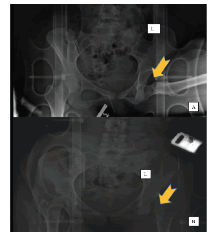
Compared to the prior imaging, a repeat Pelvic X-ray verified an obturator hip dislocation with associated iatrogenic femoral head fracture (Fig. 1A and 1B).
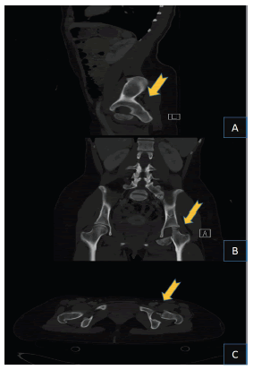
We progressed with a CT scan revealing a Pipkin classification type II. (Fig 2A-C)
It was decided an emergency open reduction of the left hip and internal femoral head fixation be performed. The patient was admitted and obtained clearance from the on-call anesthesia team. The patient was placed in a lateral position on a radiolucent operating room table, with the left hip exposed, and draped for posterior hip approach and Trochanteric flip osteotomy. The skin was incised around 15 cm from the standard posterior hip approach landmark, the gluteal maximus muscle was divided as described by the classic technique. After predrilling, a trochanteric flip osteotomy was performed.
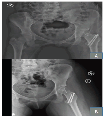
Next, the gluteal Medius was elevated with an osteotomized greater trochanter and vastus lateralis as one sleeve, preserving the piriformis muscle and a short external rotator muscle. Z-shaped capsulotomy was performed, the femoral head was exposed, and a fracture was identified. There was posterosuperior impaction of the femoral head, which was elevated using a tiny osteotome to accomplish anatomical reduction then fixed by two headless screws. Afterward, the hip joint was relocated, and the osteotomy site was fixed with two parallel 4.0 mm cannulated screws. The reduction was satisfactory with the image intensifier (Fig. 3A and 3B).
Four weeks from surgery, Enoxaparin 40 mg subcutaneously was administered along with an effective analgesic. The day following surgery, the patient began weight-bearing and range-of-motion rehabilitation. The patient was discharged home after four days on the same physiotherapy regimen for 8 weeks, with regular clinic visits. Two weeks post-surgery, the patient was examined in the clinic with a well-healed wound and normal laboratory results.
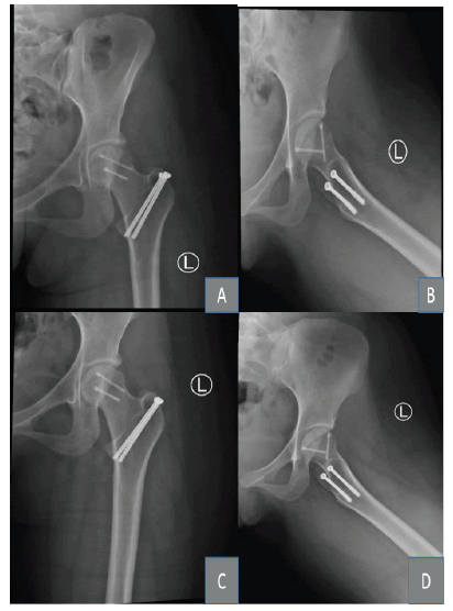
After eight weeks, she resumed her weight-bearing activities. The patient was monitored every three months for two years post-surgery, with no clinical or radiological indications of AVN or other complications (Fig 4A-D). The patient resumed her previous athletic endeavor.
Discussion
Hip dislocation resulting from trauma is a severe injury that generates major comorbidities; particularly, the length of time required to decrease the hip has a considerable impact on the chance of developing osteonecrosis of the femoral head [5].
It is an orthopedic emergency that frequently necessitates immediate reduction with close reduction procedures and adequate sedation. Close reduction is not always possible in the presence of accompanying injuries such as a femoral neck fracture, femoral head fracture, or an incarcerated bone fragment [6].
Traumatic obturator hip dislocation is an uncommon condition associated with 15%-35% superior lateral femoral head impaction but not a fracture [7]. The force applied in this position appears to be the most possible mechanism for obturator hip dislocation during forceful abduction, external rotation, and flexion of the hip [8].
In our patient’s case, a history of falling from a horse with an abducted, flexed left lower leg led to her current condition. The risk of complications, such as iatrogenic femoral head fracture, can be mitigated by an expert healthcare provider who performs a proper reduction technique.
Following a femoral head fracture, the chance of AVN is approximately 11.9%, while the risk of post-traumatic arthritis is 20% [9].
In the reported case iatrogenic femoral head fracture followed reduction of obturator hip dislocation by an ED physician, a complication caused by an improper procedure. To minimize the risk of this complication, particularly in the case of an obturator hip dislocation, close reduction required inline traction to disengage the femoral head, followed by flexion and internal rotation, which returned the limb to its original position of extension, adduction, and internal rotation [10].
Some published articles critique abduction, which may result in fractures of the femoral neck [11, 12]. Within six hours after a femoral head fracture occurs, anatomical redaction and congruent joint reduction must be performed to reduce the incidence of ANV and associated complications [13, 14].
Conclusion
Obturator hip dislocation with an iatrogenic femoral head fracture is a very uncommon injury; however, understanding the close reduction techniques, challenges, urgent reduction, and fixation within 6 hours might reduce the risk of complications.
Close observation and follow-up following the injury can result in a positive outcome.
Consent
The patient authorized the publication of this case report in writing
References
- Azar N, Yalçinkaya M, Akman YE, et al. Asymmetric bilateral traumatic dislocation of the hip joint: a case report. Eklem hastaliklari ve cerrahisi= Joint diseases & related surgery. 2010 Aug 1; 21(2):118-21. [Google Scholar]
- Ashraf T, Iraqi AA. Bilateral anterior and posterior traumatic hip dislocation. J orthop trauma. 2001; 15(5):367-8. [Google Scholar]
- Dudkiewicz I, Salai M, Horowitz S, et al. Bilateral asymmetric traumatic dislocation of the hip joints. J Trauma Acute Care Surg. 2000; 49(2):336-8. [Google Scholar]
- Phillips AM, Konchwalla A. The pathologic features and mechanism of traumatic dislocation of the hip. Clin Orthop Relat Res. 2000; 377:7-10. [Google Scholar] [CrossRef]
- Brooks RA, Ribbans WJ. Diagnosis and imaging studies of traumatic hip dislocations in the adult. Clin Orthop Relat Res(1976-2007). 2000; 377:15-23. [Google Scholar]
- K. Pfeifer, M. Leslie, K. Menn, et al. Traumatic obturator dislocation of the hip joint: About 2 cases and review of the literature. Int J Surg Case Rep. 2022 30:106983. [Google Scholar] [CrossRef]]
- Pfeifer K, et al. Imaging findings of anterior hip dislocations. Skeletal radiology. 2017; 46:723-30. [Google Scholar] [CrossRef]
- Zengui ZF, et al. Traumatic obturator dislocation of the hip joint: About 2 cases and review of the literature. Int J Surg Case Rep. 2022 30:106983. [Google Scholar] [CrossRef]
- V Giannoudis 1, G Kontakis, Z Christoforaki, et al. Bilateral simultaneous anterior obturator dislocation of the hip by an unusual mechanism—a case report. Turk J Trauma Emerg Surg. 2012; 18(5):455-7. [Google Scholar] [CrossRef]
- Walid Bouziane, Amine Machmachi, Omar Agoumi,, et al. Management, complications and clinical results of femoral head fractures. Injury. 2009; 40(12):1245-51. [Google Scholar] [CrossRef]
- Bouziane W, et al. Pure Obturator Dislocation of the Hip, a Rare Variety of Regular Dislocations, and Long-Term Clinical Outcomes. [Google Scholar] [CrossRef]
- Toms AD, Williams S, White SH. Obturator dislocation of the hip. J Bone Jt Surg. British volume. 2001; 83(1):113-5. [Google Scholar] [CrossRef]
- Polesky RE, Polesky FA. Intrapelvic dislocation of the femoral head following anterior dislocation of the hip: a case report. JBJS. 1972; 54(5):1097-8. [Google Scholar]
- Hougaard K, Thomsen PB. Traumatic posterior dislocation of the hip—prognostic factors influencing the incidence of avascular necrosis of the femoral head. Arch orthop trauma surg. 1986; 106:32-5.[Google Scholar] [CrossRef]


 Journal of Orthopaedics Trauma Surgery and Related Research a publication of Polish Society, is a peer-reviewed online journal with quaterly print on demand compilation of issues published.