Treatment of Deformations of the Anterior Thoracic Wall-Funnel and Pigeon Chest
2 Department of Biomaterials and Medical Devices Engineering, Faculty of Biomedical Engineering, Silesian University of Technology, Zabrze, Poland, Email: jan.marciniak@polsl.pl
Received: 05-Feb-2021 Accepted Date: Mar 10, 2021 ; Published: 17-Mar-2021, DOI: 10.37532/1897-2276.2021.16(1).9
This open-access article is distributed under the terms of the Creative Commons Attribution Non-Commercial License (CC BY-NC) (http://creativecommons.org/licenses/by-nc/4.0/), which permits reuse, distribution and reproduction of the article, provided that the original work is properly cited and the reuse is restricted to noncommercial purposes. For commercial reuse, contact reprints@pulsus.com
Abstract
The essence of the study was to develop a new structural form of a type series of stabilizers intended for minimally invasive treatment of deformations of the anterior thoracic wall along with the relevant surgical instruments, as well as to develop relevant diagnostic procedures and the operative technique.
Keywords
Pectus excavatum, Pectus carinatum, stabilizers
Introduction
Surgical treatment of chest deformations entails quite an extensive surgical procedure on the anterior thoracic wall. The most common defects include the funnel chest and the pigeon chest. The funnel chest also referred to as the sunken chest or concave chest (Latin: Pectus excavatum) is a congenital defect where the distal part of the sternum and the adjacent sections of ribs are dented towards the spine, with a simultaneous forward protrusion of the xiphoid process and the development of the so-called funnel. Depending on the sources, the prevalence of this defect is quoted to be at the level of 1:400 or 1:1000 of all births. What is more certain, however, is that this defect is more frequently observed in boys than girls (ratio: 3:1) [1,2].
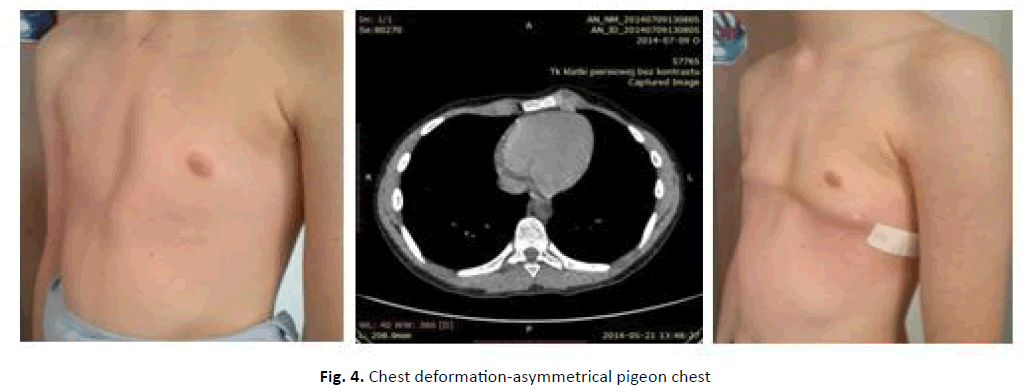
The pigeon chest (Pectus carinatum), on the other hand, is a deformation consisting of the forward protrusion of the sternum and the costal cartilages adjacent to it. The chest, protruding forward and flattened on the sides, resembles the bird’s chest, hence its name (Fig. 1). This defect is rare and-similarly to the funnel chest-it affects boys more frequently [3,4]. In 1998 Donald Nuss presented a new minimally invasive operative technique to be used in the surgery of the funnel chest, taking into account the correction of the deformation of its anterior wall, with the application of a minimally invasive technique, not requiring any bone resection [5,6].
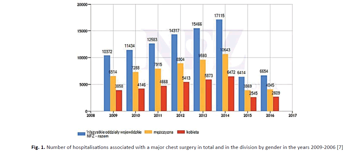
In Nuss’s method, bones are not resected nor cut. The deformation is corrected as a result of the growth of ribs along a mold based on a stabilizing plate. This refers predominantly to children, which broadens the indications for surgery in early childhood, at the age of 3 to 8. In older patients it is possible to obtain a better cosmetic result; however, in such patients, it entails considerable difficulty in distorting the deformed anterior thoracic wall, where the cartilaginous parts are already ossified. The demand for this type of surgery is illustrated in Fig. 1 [7].
While analyzing the data from the National Healthcare Fund from the years 2009-2016, it can be observed that the number of hospitalizations of patients subjected to the primary or secondary reconstruction of congenital and acquired chest deformations concerning all patients hospitalized due to diseases of the musculoskeletal system constitutes ca. 2.2%.
Objective of the Study
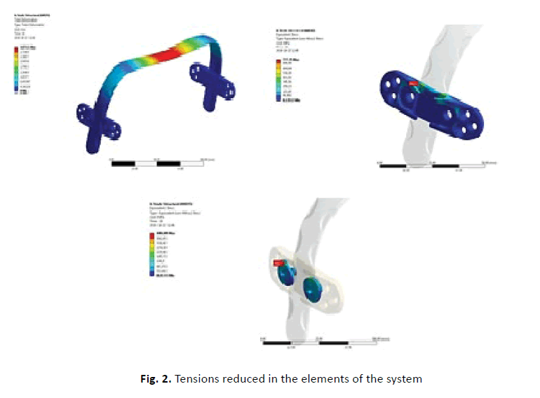
To address problems yet unsolved in this respect, as well as the current needs of the national healthcare system, an interdisciplinary study was undertaken in the Biomedical Engineering Centre of the Silesian University of Technology, within the scheme of implementing a longterm research project entitled “A New Method of Surgical Treatment of Deformations of the Anterior Thoracic Wall”. The scientific purpose of the project was to develop a new type series of stabilizers intended for the surgical treatment of the funnel and pigeon chest. The study analyzed biomechanical characteristics of the type series of individual elements of stabilizers of the state of displacement, deformation, and tension (Fig. 2) with the application of numerical and experimental methods, with the simultaneous identification of the optimal metal biomaterial. The analyses adhered to the clinical recommendations associated with the implantation technique (Fig. 3 and Fig. 4). The previous surgical experiences had pointed to the need to reduce the rigidity of the system: stabilizing plate-chest. Modification of the surface layer of the implants was also considered to enhance its corrosion resistance and biocompatibility, focusing also on the optimal sterilization method in conditions adequate to the sterilization used clinically.
The essence of the developed solution was a dimensional type series of stabilizers for surgical treatment of chest deformations in children and adolescents along with relevant surgical instruments and operative techniques. The subject matter of the study belongs to the area of interest of thoracic surgery. The new generation of stabilizers offers new possibilities. They reduce the risk of post-operative complications as they can be dedicated to personalized anthropometric and biomechanical features of an individual patient, and in doing so they can enhance the comfort of physical and mental rehabilitation of patients. The solution of the structural forms of stabilizer elements is universally useful for the surgical treatment of both the funnel chest and the pigeon chest. Other signaled solutions, including ChM [8], American patents the US No. 6 024759 [9] and US No 7 156 847 B2 [10], do not guarantee the functional properties referred to above. Our solution is protected with the patent RP P No. 222 153 [11]. It was also awarded the title “Innovator of Silesia 2013” for taking 1st place in the category “Research and Development Sector Institution” and with the Special Award of the Marshal of the Province of Silesia.
The property that makes the new generation stabilizers stand out is the fact that the plate supporting the stabilizer guarantees an even distribution of pressure onto ribs, adequately to the shape of the ribs. It does not exert point pressure upon ribs, which would cause pain and entail the need to use analgesics on a long-term basis. Four zones of attachment of the transverse plates to the ribs on each side of the supporting plate secure its stable position with the ribs and eliminate rib damage caused by the mobility of the supporting plate. This in turn reduces biomechanical and reactive complications, as well as guarantees a controlled course of the assumed therapy, which distinguishes this technique from the American solutions. The new generation stabilizers offer new possibilities of chest correction in the presence of a greater diversity of defects, reduce the risk of post-operative complications since they are adapted to personalized anthropometric and biomechanical features of the patient, increasing the comfort of their physical and mental rehabilitation.
The cognitive research from this area was carried out within the scheme of project No. N N518 415938 [12] sponsored by the National Centre for Research and Development. The prototypes of the stabilizers and adequate instruments were produced according to our documentation. The clinical trials were carried out in the Centre for Paediatrics and Oncology in Chorzów. The results of the clinical observation of 160 patients in age groups from 4 to 18 of different genders provided grounds for the final verification of the functional features of the stabilizers and the associated instruments.
Material and Method
Within the scheme of the study anthropometric measurements of chests were carried out in children aged from 4 to 18: healthy ones, and ones with deformed chests. The measurements provided the grounds for determining the features of individual elements of the stabilizers. Structural forms were developed-geometrical features of individual elements of the stabilizers intended for different chest deformations and anthropometric features of the patients, as well as mechanical properties of the biomaterial for the plates were properly selected. The biomaterial used included steel AISI 316 LVM with the chemical composition compliant with the recommendation of the standard [13], and titanium, grade 4, with the composition and properties compliant with the recommendations of the standard [14]. Furthermore, optimal conditions of surface processing of individual elements of the stabilizers were developed, taking into account the operative technique and sterilization to improve the corrosion resistance of the metal biomaterials.
Corrosion resistance tests of elements of the stabilizers [15] and the systems: stabilizers-chest models were carried out in the presence of a corrosive environment. Furthermore, the risk analysis was conducted and the implanting procedures were developed. The study also provided the foundations for the compilation of the detailed design documentation and the development of relevant surgical tools. In vitro, static and dynamic tests were performed. Based on numerical and experimental tests mechanical characteristics of the system: stabilizerribs were identified in conditions adequate to the operative technique and rehabilitation [16,17].
Results
The extensive research program provided grounds for the development of optimal parameters of the production process of the stabilizers, their biomechanical characteristics, recommendations for the operative technique, and post-operative rehabilitation of patients. The detailed design documentation of the stabilizers and associated instruments were submitted for implementation in the production and promotion of these devices in healthcare centers.
DISCUSSION
Studies regarding applied metal biomaterials, i.e. AISI 316LVM steel and, alternatively, titanium commonly used for the production of surgical implants have shown that these biomaterials are suitable for the production of the proposed form of stabilizers. Comprehensive research of corrosion resistance in the environment of in vitro and in vivo has shown that preoperative pre-bending, as well as the reactivity of the surface layer, do not initiate adverse peri-implant reactions during the treatment of deformity. The applied strengthening of biomaterials in the technological process and appropriately selected dimensional features of the stabilizers adjusted to individual anthropometric features of the patient enable the proper distribution of stress in the contact zones with the ribs, minimizing pain.
The extensive research program provided the basics for the development of optimal parameters for the production of stabilizers, biomechanical characteristics, recommendations for the surgical technique, and postoperative rehabilitation of patients. The documentation of the stabilizers and instruments was provided for the implementation of these products for production and dissemination in health care centers.
CONCLUSION
Based on the presented data, the following conclusions can be stated:
• A new method of surgical treatment of the anterior chest wall distortion was developed, as well as new types of a series of dimensioned stabilizers for the surgical treatment of funnel-shaped and chicken chests adapted to the individual anthropometric features of patients
• The biomechanical characteristics of the series of types of stabilizer elements concerning the state of displacement, deformation, and stress were analyzed using numerical and experimental methods with a simultaneous selection of the optimal metal biomaterial. The clinical recommendations related to the implantation technique were included in the analyses
• Modification of the surface layer of implants was also considered to increase its corrosion resistance and biocompatibility, also taking into account the optimal sterilization technique in conditions adequate to clinical us
• New generation stabilizers offer new possibilities. They reduce the risk of postoperative complications as they can be dedicated to the patient’s personalized anthropometric and biomechanical features, thus increasing the comfort of physical and mental rehabilitation of patients
REFERENCES
- Jaroszewski D., Notrica D., McMahon L., et al.: Current management of Pectus excavatum: a review and update of therapy and treatment recommendations. JABFM. 2010;23:230-239
- Kelly R.E., Shamberger R.C., Mellins R.B. et al.: Prospective multicenter study of surgical correction of Pectus excavatum: Design, perioperative complications, pain, and baseline pulmonary function facilitated by internet-based data collection. J. Am. Coll. Surg. 2007;205:205-216
- Abramson H., D’Agostino J., Wuscovi S.: A 5-year experience with a minimally invasive technique for pectus carinatum repair. J Pediatr. Surg. 2009;44:118-124
- Abramson H.: A minimally invasive technique to repair pectus carinatum. Preliminary report. Arch Bronconeumol. 2005;41: 349-351
- Nuss D., Kelly Jr R.E., Croitoru D.P., et al.: A 10-year review of a minimally invasive technique for the correction of pectus excavatum. J Pediat Ssur. 19981;33:545-52
- Nuss D., Kelly Jr R.E.: The minimally invasive repair of pectus excavatum. Operat Tech Thorac Cardiovascular Sur. 2014;19:324-47
- https://statystyki.nfz.gov.pl
- https://chm.eu
- The patent US nr 6024759, 2000. Method and apparatus for performing Pectus excavatum repair
- Patent US nr 7156847B2, 2007. Apparatus for the correction of chest wall deformities such as pectus carinatum and method of using the same
- Patent RP nr 222 153, 2013. Stabilizator do leczenia znieksztalcen przedniej sciany klatki piersiowej.
- Projekt badawczy nr N N518 415938. Nowa metoda leczenia operacyjnego znieksztalcen przedniej sciany klatki piersiowej”. Centrum Inzynierii Biomedy-cznej Politechniki Slaskiej, Zabrze 2013.
- Norma ISO 5832-1. Implants for surgery-Metallic materials-Part 1: Wrought stainless steel.
- Norma ISO 5832-2:2012. Implants for surgery-Metallic materials-Part 2: Unalloyed titanium.
- Walke W., Golombek K., Kajzer A., et al.: Corrosion resistance, EIS and wettability of the implants made of 316 LVM steel used in chest deformation treatment. Arch Metallur Mat. 2016;2:767-770.
- Kajzer W., Kajzer A., Gzik-Zroska B., et al.: Comparison of numerical and experimental analysis of plates used in treatment of anterior surface deformity of chest. Info Tech Biomed. 2012:319-330.
- Kajzer A., Kajzer W., Gzik-Zroska B., et al.: Experimental biomechanical assessment of plate stabilizers for treatment of pectus excavatum. Acta Bioengineer Biomechanic. 2013;15.


 Journal of Orthopaedics Trauma Surgery and Related Research a publication of Polish Society, is a peer-reviewed online journal with quaterly print on demand compilation of issues published.